Gross examination:
The thyroid may be markedly enlarged in fetuses who receive maternal thyroid stimulating antibodies via placental transfer1,2. This may occur whether the mother is or is not euthyroid following thyroidectomy or thyroid suppressive therapy (Fig 1). The fetus may be hydropic from heart failure and cachectic.
The thyroid may be absent (Fig 2)

or sometimes ectopic at the base of the tongue. For this reason, removing the neck block above (cephalad) to the hyoid bone will provide a cross section sample for histologic evaluation of ectopic thyroid tissue. A sample of the thyroid perpendicular to the trachea and including a cross section of the neck organ block often demonstrates the parathyroid as well.
Microscopic evaluation:
Colloid in follicles: Temperature modulation in utero is to a large extent controlled by the mother’s body’s temperature. The developing thyroid does accumulate iodinated protein within variable size follicles. Circulating thyroid hormone in the fetus is mostly reverse T4 which prevents overheating of the fetus in the body-temperature water bath of amniotic fluid. TSH is produced in the fetal hypothalamus and placenta at a basal rate. However, with birth, there is TSH surge that results in release of active thyroid T3 and T4 that initiates necessary thermogenesis in the fetus3. This surge occurs within the first 15 minutes. In the sheep, if a lamb is delivered but the attachment of the umbilical cord remains functional to the placenta, the externalized lamb will become hypothermic. Cutting the cord initiates the surge of TSH and epinephrine necessary to maintain body temperature outside the womb.
This histology of loss of follicular colloid in the 24 to 48 hours after birth has been documented in newborn infants at autopsy4 (Fig 3a-c).

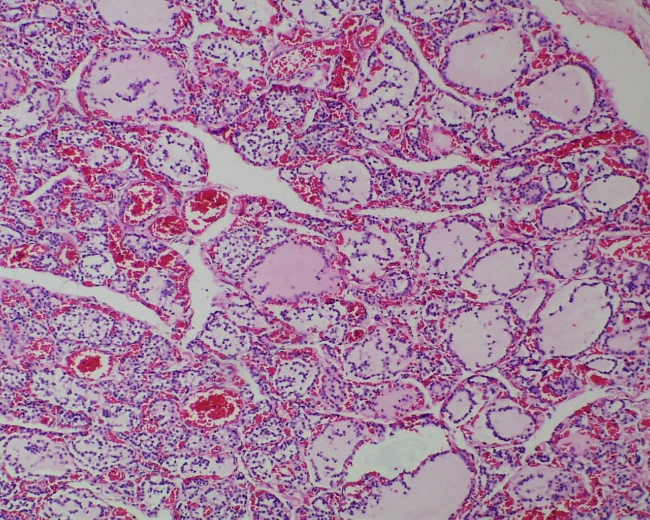
In stillborn infants the extent of colloid in the thyroid follicles and mononuclear cells in the follicles varies. Some variability may be due to gestational age differences (Fig 4a, b).


Another variable may be the effect of autolysis, but this has not been carefully correlated with postmortem retention intervals. Correlations of thyroid histology with disease in the newborn infant has been studied5. There is not a study correlating the changes in colloid with other findings in stillbirth, such as evidence of acute asphyxia or thymic involution.
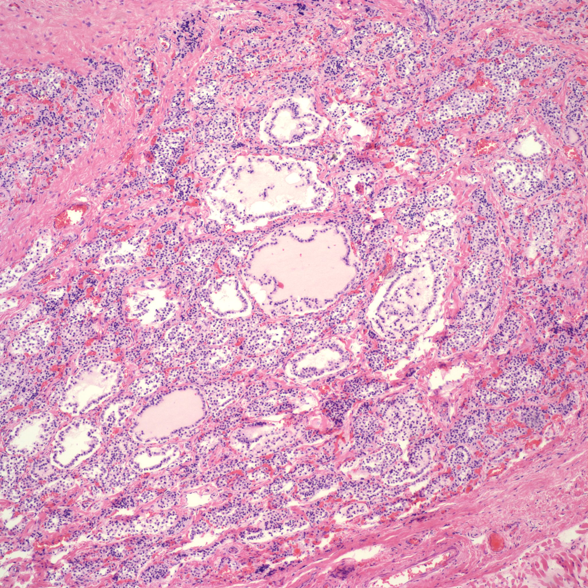
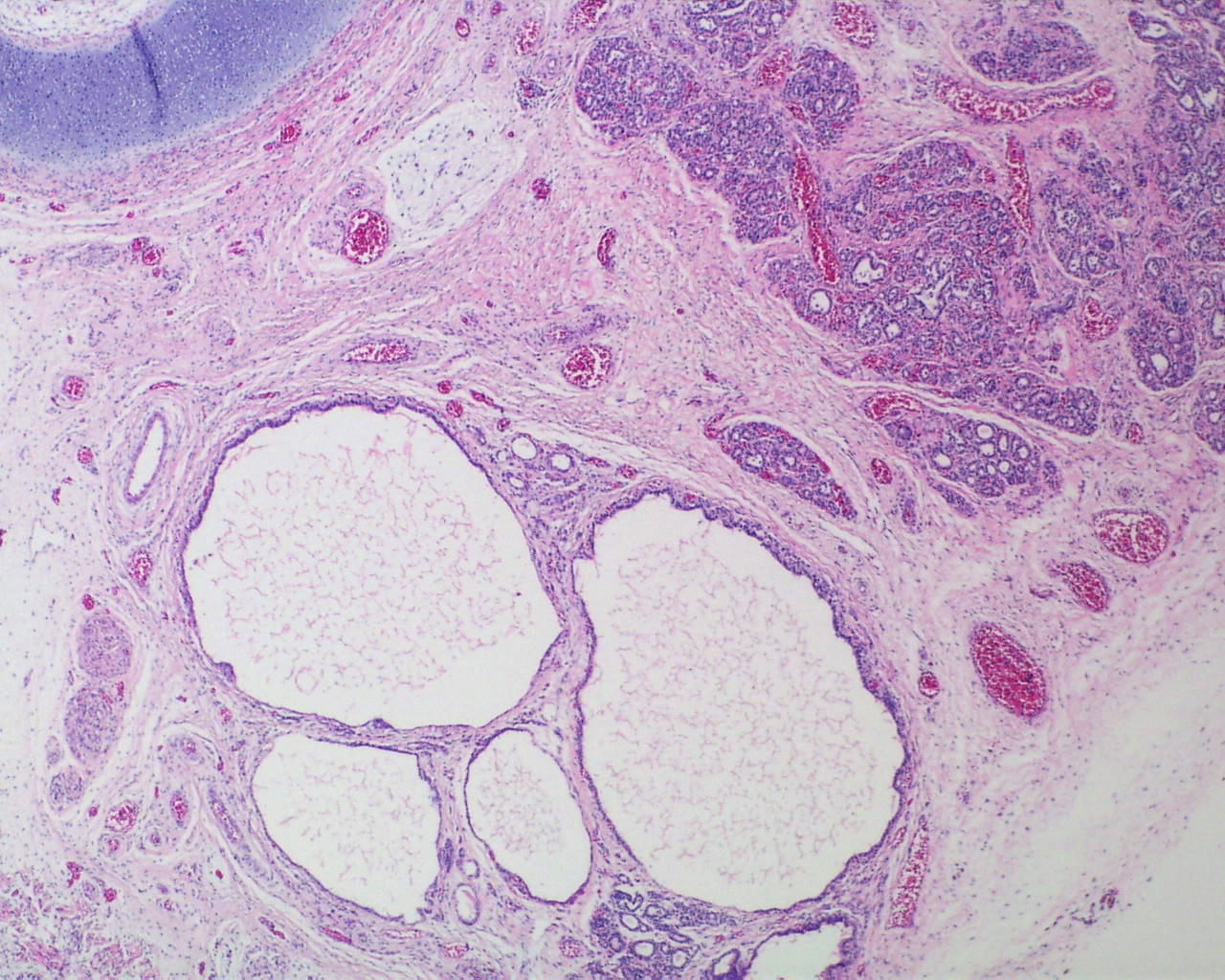
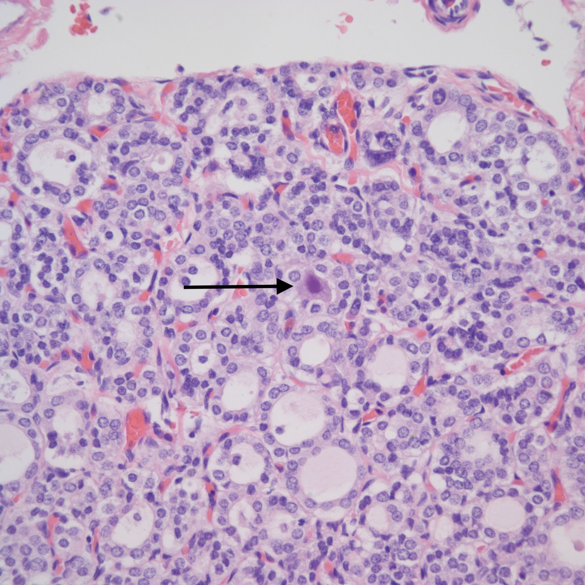
Follicular epithelium: Normally the epithelium is flat to cuboidal. However, in the presence of thyroid stimulating antibodies, there a decrease in colloid and the epithelium becomes more columnar6 (Fig 5a,b).
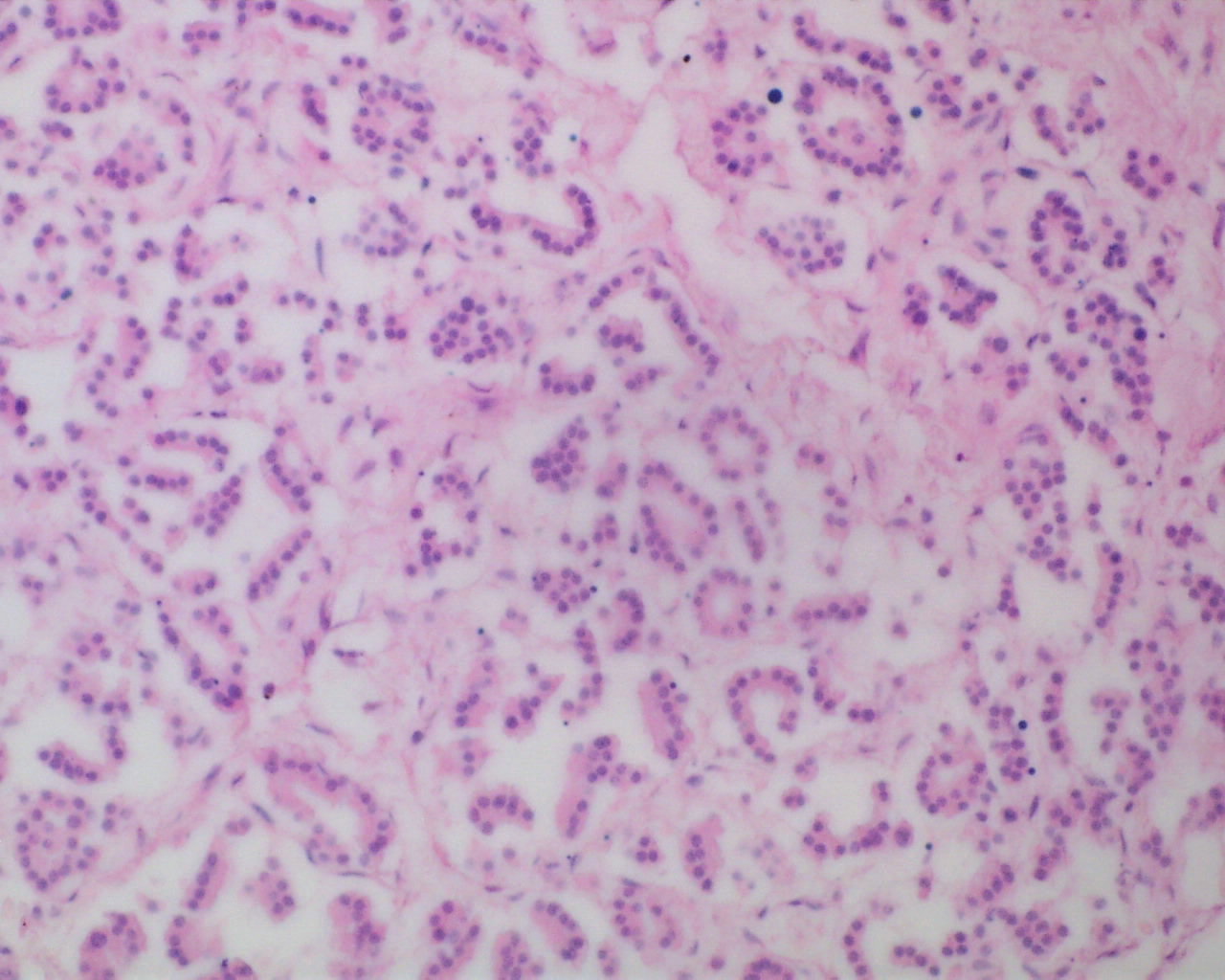
Other abnormalities: The thyroid can have focal clusters of enlarge follicles usually in the context of a maldevelopment syndrome including chromosomal trisomies. (Fig 6a, b).

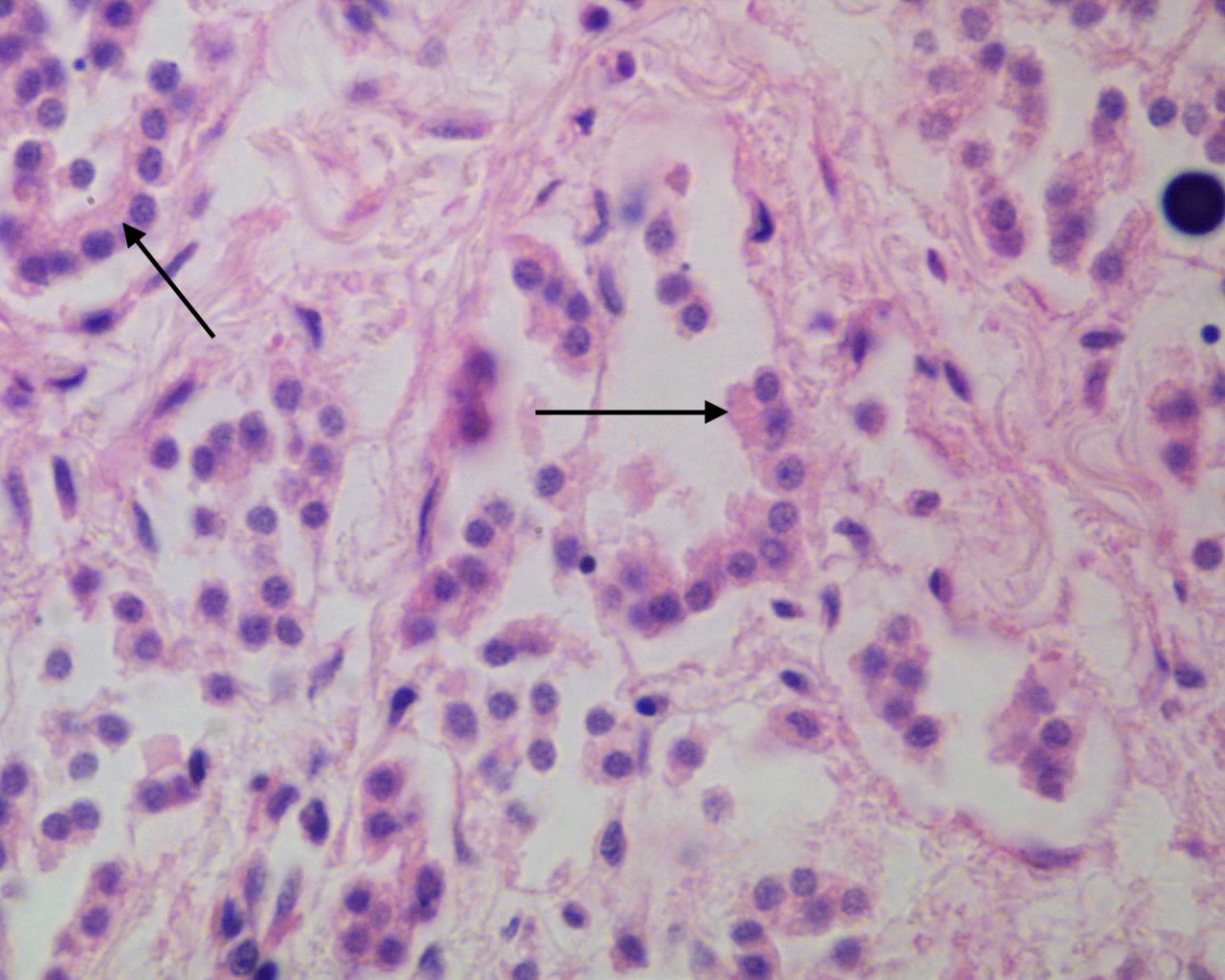
The thyroid is al tissue that may have viral inclusions in congenital Cytomegalovirus infection (Fig 7).
References:
1. Munro D, Dirmikis S, Humphries H, Smith T, Broadhead G. The role of thyroid stimulating immunoglobulins of Graves’s disease in neonatal thyrotoxicosis. Br J Obstet Gynecol 1987;85:837-43.
2. Mestman J, Manning P, Hodgeman J. Hyperthyroidism and pregnancy. Arch Intern Med 1974;134:434-9.
3. Abuid J, Stinson DA, Larsen PR. Serum triiodothyronine and thyroxine in the neonate and the acute increases in these hormones following delivery. J Clin Inv 1973;52:1195-9.
4. Sagreiya K, Emery JL. Perinatal thyroid discharge: A histological study of 1225 infant thyroids. Arch Dis Child 1970;45:746-54.
5. Larroche JC. Histological structure of the thyroid gland in the newborn. With special reference to hypotrophy, hydrops fetalis and Cesarean section delivery. Biol Neonate 1976;28:118-24.
6. Skeleton M, Gans B. Congenital thyrotoxicosis, hepatosplenomegaly and jaundice in two infants of exophthalmic mothers. Arch Dis Child 1955;30:460-4.
