Clinical History: This was a near term, 2,800 g infant. The fetal membranes ruptured while the fetal head was at 0 t0 -1 station. The infant then demonstrated 2 hours of deep variable heart rate decelerations with loss of variability one hour after they begin. For the last 30 minutes, the tracing shows progressive bradycardia “stair case of death” pattern. The infant was delivered by Cesarean section, and demonstrated a nuchal cord wrapped 3 times around the fetal neck. The neonatal resuscitation was unsuccessful.
Placental Findings: The microscopic sections from the placenta demonstrated acute inflammation in the umbilical vein in 2 of 3 samples without chorioamnionitis. The fetal membranes demonstrated meconium-laden macrophages. There were increased nucleated red blood cells in the fetal vessels.
Autopsy Findings: There were petechiae in the thymus, The lungs showed aspirated meconium. The liver showed marked congestion.
Discussion: The fetal heart rate tracing in this case is consistent with an intermittent umbilical cord compression pattern that eventually led to fetal acidosis and death. At Cesarean section, the obstetrician reported the cord wrapped three times around the neck. The evidence of umbilical cord compression in the fetal heart tracing followed the rupture of the membranes. This sequence might be explained by an occult prolapse of the cord along the fetal head because of the position of the free cord due to the nuchal wrapping. The other possibility is that the distance between the cord wrapping and the placenta was short making the cord susceptible to occlusion from slight torsion or from downward traction. The small number of neutrophils in the cord without other evidence of ascending inflammation is consistent with direct injury to the cord. There was mild evidence of meconium aspiration that along with intrathoracic petechiae is evidence of acute asphyxia with gasping respiration. The liver congestion and prolonged clinical course suggest some prolonged acidosis and heart failure must also have developed. The nucleated red blood cells are of moot significance since the mother was O+ blood type with anti-A antibodies on direct Coombs test.

Fig 1) This fetal heart rate tracing on the right side shows a deep variable deceleration. The beat to beat variability is normal.
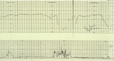
Fig 2) This later tracing shows 2 long deep variable decelerations, and loss of normal beat-to-beat variability.

Fig 3) This even later tracing shows progressive bradycardia.

Fig 4) There are sparse subintimal neutrophils in the umbilical artery

Fig 5) This section of chorion shows the absence of neutrophils. There are meconium macrophages but they do not display well.

Fig 6) There were increased nucleated red blood cells seen here in the umbilical vein.
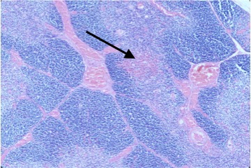
Fig 7) The arrow points to a petechial hemorrhage in the thymus.

Fig 8) The arrow points to squames from aspirated vernix and meconium. The pigment does not display well.
