Introduction
Asphyxia in modern English is another term for suffocation, the failure of respiratory gas exchange. In the fetus, this suffocation occurs because of a failure of gas exchange between the fetal blood in the placenta and the maternal uterine blood perfusing the placenta. The effect of asphyxia is a lowering of tissue oxygen and carbon dioxide (respiratory acidosis) and with time a metabolic acidosis from anaerobic metabolism. Such asphyxia can be complete, partial or intermittent. In experimental models in the monkey and sheep precise durations and degrees of umbilical cord blood flow occlusion have been correlated with the timing of fetal hypoxia and acidosis, as well as with the resultant changes in fetal heart rate. Such studies have also defined parameters that lead to fetal organ injury, particularly brain necrosis, and to fetal death.
While the fetus is still in the uterus, asphyxia is usually diagnosed by inferences from the fetal heart rate tracing. Late fetal heart rate decelerations occur after contractions in which uterine blood flow to the placenta is reduced. The carotid body responds to the transient hypoxia by vagal slowing of the heart rate. Deep variable fetal heart rate decelerations are usually due to occlusion of umbilical cord blood flow. Decreased fetal heart rate beat to beat variability indicates decreased central nervous system feed to heart rate communication. Bradycardia often indicates severe cardiac under-perfusion. The problem with fetal heart rate recording is that not all the variables that might predict severe brain injury or death are summed up by the heart rate changes. As Dr. Ron Myers discovered, the prior state of the infant may determine the degree of brain injury for a given subsequent asphyxial event. We will review these studies later.
From a pathologist’s point of view, a major defect of the experimental studies is that they use an inflatable balloon around the cord to produce asphyxia. The studies do not elucidate the natural causes of fetal asphyxia. Asphyxia could be due to structural changes in the placenta that reduce the amount of oxygen that can be extracted between contractions, or it might be due to a mechanical compromise of the cord, or might be due to spasm of the umbilical vessels. Ideally knowing the cause of asphyxia quickly in relationship to fetal heart rate changes might lead to better assessment of treatment options to alleviate the asphyxia prior to resorting to urgent Cesarean delivery, and better define when emergency delivery is truly needed to save the fetus from injury or death. As we will see, a major proportion of stillbirths have evidence of having died from asphyxia Progress in prevention of stillbirth will require the ability to identify the mechanism of that potential asphyxia before fetal death occurs.
I will first review the historical development of our understanding of fetal asphyxia. Then I will present the evidence from autopsy observations that asphyxia is a major cause of stillbirth. Finally I will discuss mechanisms of fetal asphyxia and evidence about the type and duration of intrauterine asphyxia, derived from examination of the placenta and umbilical cord.
The dictionary argument against the term “intrauterine asphyxia”
The Greek root of asphyxia means pulseless. The Oxford English Dictionary cites this meaning as early as 1706 attributed to Phillips: “Asphyxia which is the highest degree of swooning next to death”. In 1731, Bailey used it in a similar manner, “in some cases where it stops for a time”. This use seems to correspond to our current notion of cardiac arrest. This usage seems to attest to some spontaneous survival; otherwise it would be the equivalent of death. The restriction of the term to suffocation came later. The first citation is a 1778 book by Brand entitled “The cure of Asphyxia or apparent death by drowning”. This is the use that is common today. This deviation from the original root bothered Dr. DeLee so much that in his textbook “Principles and Practice of Obstetrics” he proposed that the term be changed to follow the Greek root for privation of air and be termed anaerosis. The term never caught on.
The other dictionary argument quoting directly from a debate about the term on the listserve of the Society for Pediatric Pathology that the dictionary says “that asphyxia is the interference with breathing in air”. I would argue that changing that definition to interference with respiration, that is with the exchange of carbon dioxide and oxygen with the environment, is a more reasonable definition. When fish die in air, I have no trouble thinking, that the fish suffocated. I do not have to think that the fish died of piscine hypoxia. here is another reason that I stick with the term intrauterine asphyxia. Asphyxia points to an active agent rather than a passive state. Something has to suffocate you. Fetal hypoxia or to use another euphemism “fetal intolerance to labor” both suggest a passive event. Uppermost in this section will be the search for the causes and possible cures of intrauterine asphyxia. So without further apology, we will examine the nature of intrauterine asphyxia.
Aside #2 Clarification of fetal respiration
A sometime misunderstood point: Intrauterine asphyxia is an interruption of the exchange of oxygen and carbon dioxide between the mother and fetus via the fetal and maternal circulation of the placenta. Gas exchange takes place with the atmosphere only in that the mother transports oxygen from the air to the fetus, and the carbon dioxide from the fetus to the air. Intrauterine asphyxia has absolutely nothing to do with the airway of the fetus. Wrapping the umbilical cord around the neck of the fetus, a nuchal cord, even if tight does not directly interfere with fetal respiration. Fetal respiration is happening in the placenta, not in the fetal lungs. The fetus does make the motions of shallow breathing with a small transfer of amniotic fluid, but this is not for respiration.
John William Little (1810-1894)
John William Little was an orthopedic surgeon who had mastered the technique of tenotomy (cutting the tendon) to relieve the shortened muscles of spastic limbs [1]. Dr. Little did not comment on the role of fetal asphyxia as a cause of ankylosis (fixed rigid joint) in his 1843 monograph on ankylosis1, but his subsequent fame must have provided him with a busy referral practice of caring for such children. An individual obstetrician or pediatrician would not have seen many or any cases of such spastic paralysis and ankylosis, but Dr. Little saw in his practice approximately 200 cases over 20 years. During that time, he recorded detailed histories from the mothers of these patients and from this unique perspective found the common thread that obstetrical difficulties, often with periods of postpartum fetal apnea, were the antecedents of the spastic joints. He made the analogy of birth asphyxia with brain injury from “suffocation in later life from drowning, when the air passages are suddenly and forcibly obstructed, suffocation also from inhalation of certain gases which exclude oxygen from the lungs,”. He presented his observations and deductions to the London Obstetrical Society in 1862. Excerpts from those transactions can be read in the attached text fragments Citations 1-9. Today many of his cases would be classified as cerebral palsy, sometimes still called Little’s Disease.
Dr. Little stated his case very forcibly. He was aware of some specific causes of intrauterine asphyxia including cord compression and placental separation. He used the term suspended animation basically as a synonym of asphyxia. He commented on oxygen and was aware of its importance, but seems to relate the consequence of lack of respiration to carboxylation of blood. His remarkable observations followed from a strong grasp of the contemporary basic science, a focus on a very specific diagnosis namely limb spasticity that required his intervention, and the curiosity to ask the mother about the events in the child’s birth and life, and to record the answers.
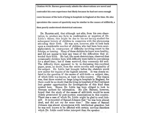
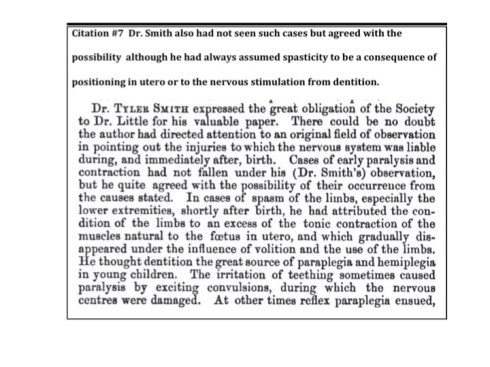
How did the obstetricians of the day react to his presentation? They reacted with understandable skepticism, but were impressed by the evidence. See citations 6 and 7 . As individual obstetricians they could not personally have seen the large number of cases of intrauterine asphyxia that Little had referred to him. They tended to confuse association with cause and effect, for example seizures as the cause rather than consequence of asphyxia.
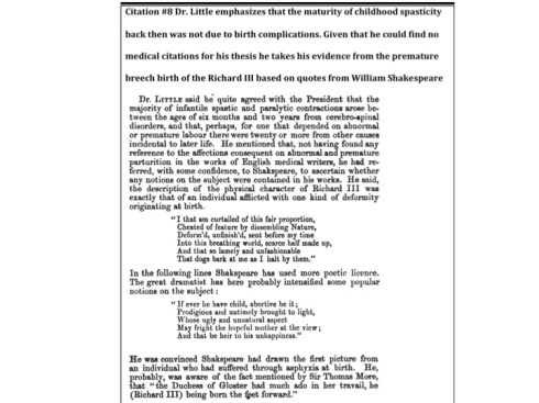
Little’s individual case reports are a fascinating window on medical thinking of the time. For example, the mother having a fright is often mentioned as a cause of poor obstetrical outcome. As today, it was recognized that many of the children with spastic paralysis also suffered from mental delay, seizures, and feeding difficulty. His cases included the coexistence of severe motor disorders with normal intellect, a condition poignantly recorded in the book and movie, “My Left Foot”. Case examples are presented in Citation 9. It is perhaps ironic that the title of a paper in the American Journal of Obstetrics and Gynecology published in October 2010 “Adverse obstetric events are associated with significant risk of cerebral palsy”2 reflects that of Little’s 1862 paper. Of course in 1862 there were fewer options for avoiding obstetrical complications.
- Little JW. On Ankylosis or Stiff-joint. London: Longman, Brown, Green and Longmans; 1843.
- Gilbert WM, Jacoby BN, Xing G, Danielsen B, Smith LH. Adverse obstetric events are associated with significant risk of cerebral palsy. Am J Obstet Gynecol 2010;203:328 e1-5.
Citations from Little’s On the influence of abnormal parturition, difficult labours, premature birth, and asphyxia neonat orum, on the mental and physical condition of the child, especially in relation to deformities.
Citation 1. The damage to the fetus at birth follows general pathologic principles
Little Citation 1-3Little Citation 1-3

Citation #2 Apnea at birth, not always innocuous

Citation #3 Recognition of the role of uterine contraction