The primate model of perinatal asphyxia
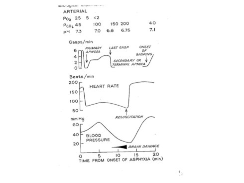
The study of perinatal asphyxia was advanced by a model in newly delivered monkeys. Immediately after delivery before breathing could be initiated, the umbilical cord was clamped, and the newborn head was immersed in a plastic bag full of warm saline. These studies while appearing heartless established the pattern of infant response and a basic vocabulary of perinatal asphyxia necessary to understand and prevent brain injury in newborn infants. Early in my pathology career I found a book about these studies in a monograph by G.S. Dawes who published in the early 1960’s. An example graph from one of these monkey’s as published by Dawes can be seen in figure 1 (Dawes 1968). The graph defined the conditions that cause primary and secondary apnea in the infant monkey, and most importantly they demonstrated that after approximately 14 minutes of not being able to breath the monkeys began to develop cardiovascular collapse and brain injury. In a sudden acute obstetrical emergency this type of research supported the urgency of intervention within that 15 minute of window.
These studies provided other valuable insights. An asphyxiated infant who doesn’t try to breath because of primary apnea is unlikely to have brain injury and may start breathing on his own with deep gasping after a short delay. On the other hand an infant in secondary apnea needs resuscitation as soon as possible to avoid brain injury and is unlikely to spontaneously recover once gasping has ceased. Resuscitation of apneic newborn is now the standard of care, and if available neonatologists rush to the scene of delivery of an infant who may have suffered intrauterine asphyxia.
A major initiative in the study of infant asphyxia based on this pioneering monkey studies was forming in the late 1950’s. A conference was held at the University of Puerto Rico on Asphyxia Neonatorum, brain damage and impairment of learning. The conference was held under the auspices of the National Institute of Neurological Diseases and Blindness. At the concluding session of the meeting Dr. Pierce Bailey who was director of that institute announced that since the formation of the institute in 1951 he realized the importance of research into neurologic deficits that occur early in life. In 1955 he gathered an ad hoc committee of experts to plan a large multi-institutional collaborative clinical study. As an obstetrical pathologist, I am very familiar with this perinatal collaborative study that resulted in many well-known pathology publications including those on the placenta by Dr. Naeye(Naeye 1992), on the autopsies by Fujikura and Froehlich(Fujikura and Froelich 1972), and the neuropathology by Drs. Gilles, Leviton and Dooling(Gilles, Leviton et al. 1983).
I did not know until I read the publication of this symposium and Dr. Bailey’s address, that the primate studies done by Dr. Ronald E. Myers and colleagues were also part of this grand plan. That under the guidance of Dr. Windle with collaboration of the University of Puerto Rico had established a breeding colony of Rhesus monkeys at Cayo Santiago where the monkeys were living and reproducing successfully. The site had laboratories and an operating room. At the end of the conference, there was a unanimous resolution passed that established “the monkey as a standard animal for research in the area covered by this conference.” The researchers named for the project at that time were Dr. Ramirez de Arellano, Mr. Smart, and Mr. Altman.
After this point, I am not sure what progressed but Dr. Ronald E. Myers eventually made extraordinary use of this opportunity. I do not know much personal information about him despite the fact that his wife Gabriella DeCourten Myers, a neuropathologist, was as a colleague with me at the University of Cincinnati, and I had the pleasure of meeting this very modest quiet man. Most of what I know I have discovered from his published papers. I did ask him once what happened to the visceral organs and the placentas from the monkeys in his studies. The placentas were given to Dr. Maurice Panigel, but the organs had been stored and he would see if they were still available now 20 years later. They were not.
I was very surprised to find out that Dr. Myers’s early studies included a seminal paper on the split brain of the cat with Dr. Sperry who went on to win the Noble prize for his work on the split brain, that is the brain with a separated corpus callosum that isolated the right hemisphere of the brain from the left(Myers and Sperry 1958). Dr. Myers extended this work to the chimpanzee.
Dr. Myers became the leader of the research done at the National Institute of Health monkey colony in Puerto Rico. His papers tell the story of a critical discovery about intrauterine asphyxia that I don’t think is fully appreciated by obstetricians today.
References:
Dawes, G. (1968). Foetal and Neonatal Physiology. Chicago, IL, Year Book Medical Publishers, Inc.
Fujikura, T. and L. Froelich (1972). “Organ-weight/brain -weight ratios as a parameter of prenatal growth: A balanced growth theory of visceras.” Am J Obstet Gynecol 112: 896-902.
Gilles, F. H., et al. (1983). The Developing Human Brain: Growth and Epidemiologic Neuropathology. Boston, John Wright . PSG Inc.
Myers, R. E. and R. W. Sperry (1958). “Interhemispheric communication through the corpus callosum: mnemonic carry-over between the hemispheres.” AMA Arch Neurol Psychiatry 80(3): 298-303.
Naeye, R. L. (1992). Disorders of the Placenta, Fetus and Neonate, Diagnosis and Clinical Significance. St. Louis, Mosby Year Book.
This is the figure from Dr. Dawes monograph that shows the typical changes in the physiological parameters of a newborn monkey that is asphyxiated immediately at birth.