Cord Width
Width of the cord:
The cross-sectional area of the cord in life as measured by ultrasound is wider because of distended umbilical blood vessels than the diameter measured in the pathology laboratory because the blood vessels are full in in vivo1. Measured by ultrasound, the umbilical cord diameter increases from approx. 0.3 cm at eight weeks of gestation to 0.5 cm at 15 weeks2,3. An ultrasound study measuring cord thickness at the umbilicus found a direct relationship with fetal size until 32 weeks of gestation, but not after that gestation4. Similar results were found with the cord diameter measured in pathology, with the diameter plateauing around 1 cm at 28 weeks of gestation Fig (1). 1
Table 1
Umbilical cord diameter percentiles and mean and standard deviation of the cohort
(N . 497). UCD was calculated using the formula: UCD . 2 O(weight/p length).
The substitution of weight in place of volume was used after showing that umbilical
cord volume was similar to weight (p < 0.001; r . 0.997; y . 1.071x 0.644).
Gestational age Umbilical cord diameter (cm)
(completed weeks) 10th percentile 50th percentile 90th percentile n Mean Standard deviation
18 0.36 0.46 0.61 4 0.53 0.08
19 0.43 0.54 0 .68 10 0.51 0.12
20 0.50 0.60 0.73 8 0.72 0.30
21 0.56 0.67 0.79 9 0.61 0.14
22 0.62 0.72 0.84 17 0.69 0.09
23 0.67 0.78 0.88 14 0.75 0.20
24 0.72 0.83 0.93 18 0.84 0.13
25 0.77 0.87 0.97 9 0.86 0.12
26 0.80 0.91 1.01 9 0.93 0.13
27 0.83 0.94 1.05 7 0.93 0.09
28 0.85 0.97 1.09 4 1.04 0.06
29 0.87 1.00 1.12 9 0.92 0.16
30 0.88 1.02 1.15 16 1.04 0.18
31 0.88 1.03 1.18 13 1.06 0.16
32 0.88 1.05 1.20 10 1.04 0.14
33 0.88 1.05 1.22 16 1.06 0.20
34 0.87 1.06 1.23 23 1.03 0.24
35 0.86 1.06 1.24 17 0.97 0.13
36 0.85 1.05 1.25 33 1.03 0.17
37 0.84 1.05 1.24 40 1.02 0.14
38 0.83 1.03 1.23 54 1.04 0.18
39 0.82 1.02 1.22 58 1.01 0.15
40 0.81 1.00 1.19 59 1.04 0.14
41 0.80 0.98 1.16 40 1.05 0.17
Proctor LK,Fitxgerals B, Whittle WL et al. Placenta (2013) 34:62-66
Diameter growth
It is not surprising that the cord thickness, like the length, grows with the fetus. However, the understanding of this process is not complete. Growth could be controlled by blood pressure, nutrient delivery, or release of growth factors in Wharton’s jelly.
One study also looked at histological sections and found that there was a disproportionate increase in umbilical arterial wall thickness in thick cords and a relative decrease in Wharton’s jelly in thin cords1. The decrease in Wharton’s jelly in the large cords may have been biased by the tissue processing that reduced overall diameter. The arterial muscle may be more resistant to shrinking than the more fluid Wharton’s jelly. However, if true, this finding would imply that thick cords may be associated with increased fetal blood flow or pressure.
The mechanism of umbilical cord respiration or nutrition must either depend on direct diffusion from amniotic fluid or diffusion from the umbilical vessels, or on both. That molecular exchange takes place across Wharton’s jelly is evident in cases of funisitis or with meconium macrophages. The level of oxygen diffusion is suggested by the crescentic volume of necrosis around a thrombosed umbilical artery. The growth in thickness may be dependent on the availability of oxygen and nutrients, in a manner analogous to the Pedersen hypothesis that substrate availability drives excess growth in infants of diabetic mothers. Likely growth in width is also controlled by release of bound growth factors that likely control longitudinal and diameter growth.
Cord edema:
Wide cords often appear edematous with translucent areas, and there is direct evidence in most of excess water content. A study using a freeze dry technique demonstrated that thick cords had more water content (>92%). Rarely some thin cords that appeared edematous had similar water content5. They found that microscopically edematous cords had more large open spaces than normal diameter cords and that in some specimens, the spaces extended into the vascular smooth muscle. These spaces can be seen easily on routine microscopy, and even in normal width umbilical cords, potential spaces distend with blood when there is hemorrhage. An SEM study of the collagen fiber skeleton of Wharton’s jelly demonstrates the lattice arrangement that may underlie these spaces6. This lattice appearance was confirmed in an immunohistochemical and ultrastructural study7. This study also documented the progression over gestation of the maturation of stromal cells toward myofibroblasts which may explain the distensibility of the spaces.
The potential of these spaces to hold free water may have functional significance, for example if the cord is compressed, the water in that area may escape along the cord preventing vascular compression from being transmitted through the jelly. If those spaces are already filled with fluid, this mechanism may not be successful.

Figure 1Typical reticular collagen (pink) with spaces filled with glycoaminoglycan gel in Wharton’s Jelly. (H&E)
There is little known about water balance in Wharton’s jelly, but like other proteoglycan matrices in cartilage or the cornea, the tissue is maintained without capillary exchange. As there is no capillary bed in the cord, Starlings “law” which relates post capillary tissue and osmotic pressure to edema, does not apply. In the cord passive edema could be related to fluid pressure driving water from the umbilical vessels or from osmotic pressure from the gel drawing water in from the vessels or amniotic fluid. An in vitro study of the perfused umbilical cord, found intravascular vascular endothelial growth factor produced edema in the wall of the umbilical vein8. This result suggests that increased endothelial permeability over time might contribute to edema of Wharton’s jelly. In pre-eclampsia this is a loss of hyaluronic acid compared to sulfated glycoaminoglycans9, but it is unclear how such changes might affect the diameter. There is little information on how the cord maintains water balance, or on other processes that can overwhelm the balance.
One paper cited an article that Wharton’s jelly is a thixotropic gel but this was not supported in the paper cited5.
Thick umbilical cords:
Normal umbilical cord width increases with gestation. In the third trimester, cords above the tenth percentile are wider than 1.25 cm1. In a study of 100 thick umbilical cords compared to 100 random controls, the thick cords were significantly associated with Rhesus iso(allo)immunization (15/50), abruptio placenta (15/24), macerated stillbirths (19/25), and 7 diabetic infants (7/17) 10. The authors commented that the abruption cases were mostly stillborn, but they did not specify fetal weights or the presence of fetal hydrops. Since the cord width is correlated with birthweight, they are also correlated with macrosomia 11. One study found starting at 24 weeks of gestation that the umbilical cords of fetuses of diabetic mothers were thick, and had increased Wharton’s jelly (determined by ultrasound subtraction of the vessel diameters from the entire diameter of the cord) compared to controls, yet the infants were not macroscomic12.
Early in gestation thick cords are a potential marker of aneuploidy3,13.
Thin umbilical cords:
A thin umbilical cord correlates with low placenta and low birthweight,1,14,15. In one study intrauterine growth restriction, there was a decrease in the cross-sectional area of not only Wharton’s jelly but also the umbilical vein and its lumen15. In cases of growth retardation with abnormal end diastolic flow on Doppler studies, there is also a reduction in arterial area and wall thickness in the cord. The consensus criteria from the Society for the Study of Pediatric Pathology is that umbilical cord diameters of less than 0.8 cm in term infants are a feature of maternal vascular malperfusion16. This does not imply that thin cords are specific for that cause of restricted growth. The difference in cord diameters can be striking when examining a twin placenta with birth weight discordance. Thin cords have also been associated with single umbilical artery (further evidence that arterial muscle is an important component of cord width) and with marginal umbilical cord insertion. This latter association at P<.02 is unexplained.
There is insufficient evidence to implicate a narrow cord per se as harmful, but a decreased ability to protect the cord vessels from compression cannot be excluded. A study of 439 fetuses 8-15 weeks of gestation found that small cords included cases of fetal demise or subsequent development of preeclampsia but did not reach statistical significance2. The authors note that cord diameter at this gestation is composed mainly of the fetal blood vessels with little Wharton’s jelly.
Massive umbilical cord edema:
Leakage of urine into Wharton’s jelly from a patent urachus has been reported multiple times17-19. The cord is generally several times the normal cord diameter and wider toward the fetus. The infant may not have other pathology beyond the patent urachus, but the lesion may also occur in the context of distal urinary tract obstruction. A non-patent urachal remnant is a common finding at the fetal end of the umbilical cord between the two umbilical arteries.
.
Potential research into cord width:
Clinical: Wharton’s jelly composition may be important in protecting the umbilical blood flow if the cord is twisted, compressed, or kinked. A thin cord in fetal growth restriction may make the cord more vulnerable to fetal hypoxia and deep variable FHR decelerations. The association of thick cords with stillbirth is not just with macrosomia but occurs in my experience with unexplained stillbirth. With a sufficiently large database, the umbilical cord diameter on prenatal ultrasound could be evaluated as a risk factor for unexpected stillbirth. A question not addressed is does discordance between cord width expected for fetal size and the actual cord width have any significance.
.
Basic: The basic science of water balance in Wharton’s jelly and of its gel properties might provide a better understanding of its role in the structure and function of the umbilical cord. Hypothetically a change in Wharton jelly such as increasing water content or a change in glycosaminoglycan composition could alter the cord’s flexibility and its protection of umbilical blood flow. If Wharton’s jelly is a thixotropic gel (meaning it becomes less viscous when a stress is applied), how does this effect the response to longitudinal tension and torsion, and the size and shape of the cord? In vitro studies of thick and thin cords, including those from stillbirths, with various manipulations could further elucidate the role of Wharton’s jelly in fetal well-being.
Practical pathology:
1) The diameter of the cord is not perfectly constant, but an estimate by measuring the middle range diameter is usually sufficient. One study found that weight of the cord was proportional to volume and could be used, along with the length, to calculate a more accurate diameter20. Fortunately, this is degree of accuracy is not required in routine pathological examination.
2) Marked focal widening should be examined by the pathologist and appropriately sampled. Patent urachus is rare, but marked widening of the cord more toward the fetal end with only edema on cut-section requires the exclusion of that diagnosis. If it is suspected, the nursery should be notified. Even more rarely, a proximal thickening is due to gastrointestinal tissue from the omphalomesenteric remnant (Meckel diverticulum) which also needs prompt diagnosis and notification of the nursery21,22. Other causes of focal thickening will be discussed elsewhere.
3) If the placenta in pathology does not have identification of the umbilical cords in discordant birthweight, monochorionic twins, the discordant cord diameters may identify which placental area belongs to which twin.
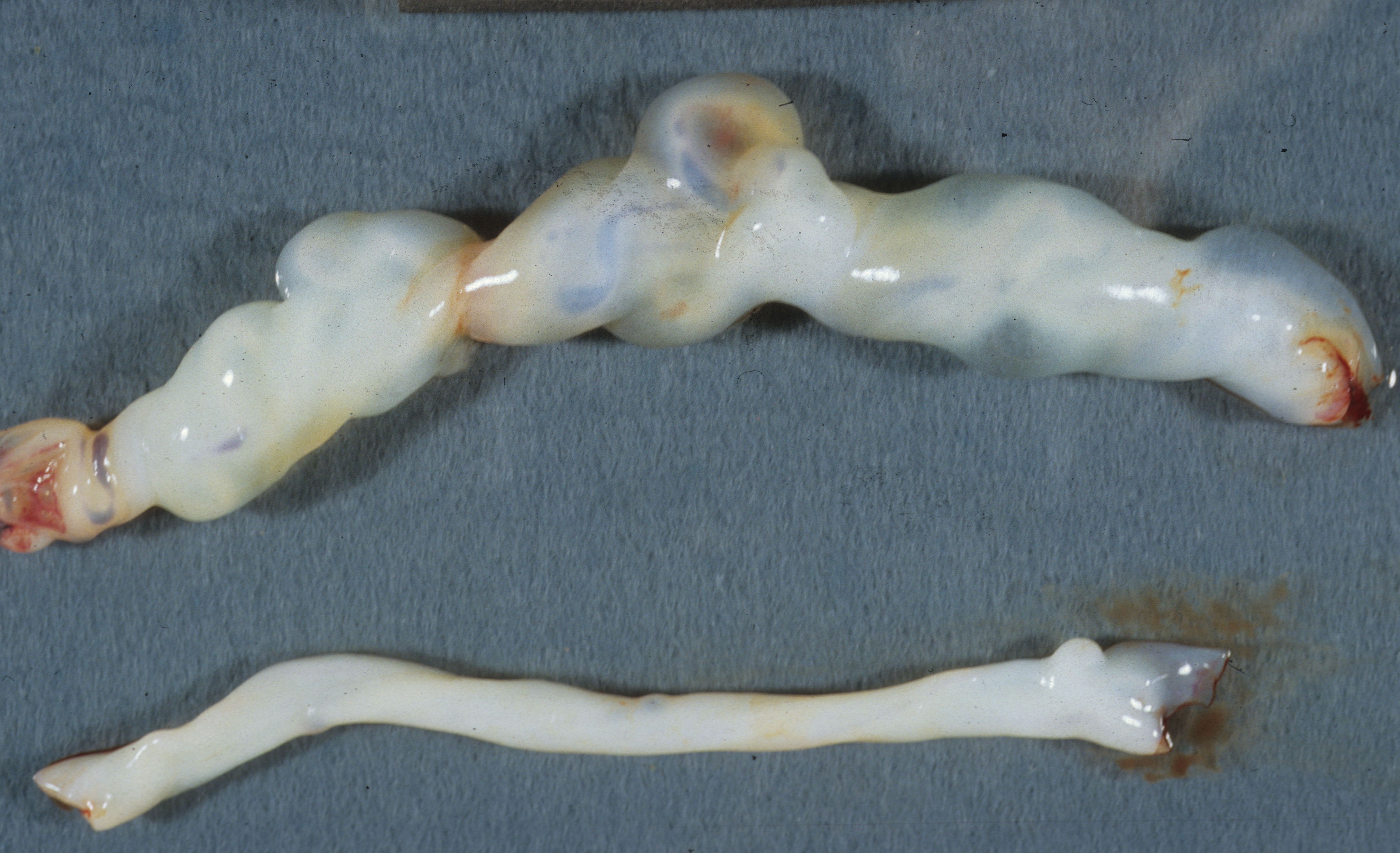
Figure 2 Umbilical cords from discordant twins
References:
1. Proctor LK, Fitzgerald B, Whittle WL, et al. Umbilical cord diameter percentile curves and their correlation to birth weight and placental pathology. Placenta 2013;34:62-6.
2. Ghezzi F, Raio L, Di Naro E, et al. First-trimester sonographic umbilical cord diameter and the growth of the human embryo. Ultrasound Obstet Gynecol 2001;18:348-51.
3. Ghezzi F, Raio L, Di Naro E, Franchi M, Buttarelli M, Schneider H. First-trimester umbilical cord diameter: a novel marker of fetal aneuploidy. Ultrasound Obstet Gynecol 2002;19:235-9.
4. Ghezzi F, Raio L, Di Naro E, Franchi M, Balestreri D, D’Addario V. Nomogram of Wharton’s jelly as depicted in the sonographic cross section of the umbilical cord. Ultrasound Obstet Gynecol 2001;18:121-5.
5. Scott JM, Wilkinson R. Further studies on the umbilical cord and its water content. J Clin Pathol 1978;31:944-8.
6. Vizza E, Correr S, Goranova V, et al. The collagen skeleton of the human umbilical cord at term. A scanning electron microscopy study after 2N-NaOH maceration. Reprod Fertil Dev 1996;8:885-94.
7. Nanaev AK, Kohnen G, Milovanov AP, Domogatsky SP, Kaufmann P. Stromal differentiation and architecture of the human umbilical cord. Placenta 1997;18:53-64.
8. Infanger M, Grosse J, Westphal K, et al. Vascular endothelial growth factor induces extracellular matrix proteins and osteopontin in the umbilical artery. Ann Vasc Surg 2008;22:273-84.
9. Romanowicz L, Bankowski E, Jaworski S. The activities of some glycosaminoglycan-degrading enzymes in the wall of the umbilical cord artery and their alteration in edema, proteinuria, hypertension (EPH)-gestosis. Clin Chem Lab Med 1999;37:417-21.
10. Coulter JB, Scott JM, Jordan MM. Oedema of the umbilical cord and respiratory distress in the newborn. Br J Obstet Gynaecol 1975;82:453-9.
11. Cromi A, Ghezzi F, Di Naro E, Siesto G, Bergamini V, Raio L. Large cross-sectional area of the umbilical cord as a predictor of fetal macrosomia. Ultrasound Obstet Gynecol 2007;30:861-6.
12. Weissman A, Jakobi P. Sonographic measurements of the umbilical cord in pregnancies complicated by gestational diabetes. J Ultrasound Med 1997;16:691-4.
13. Axt-Fliedner R, Schwarze A, Kreiselmaier P, Krapp M, Smrcek J, Diedrich K. Umbilical cord diameter at 11-14 weeks of gestation: relationship to nuchal translucency, ductus venous blood flow and chromosomal defects. Fetal Diagn Ther 2006;21:390-5.
14. Raio L, Ghezzi F, Di Naro E, et al. Prenatal diagnosis of a lean umbilical cord: a simple marker for the fetus at risk of being small for gestational age at birth. Ultrasound Obstet Gynecol 1999;13:176-80.
15. Bruch JF, Sibony O, Benali K, Challier JC, Blot P, Nessmann C. Computerized microscope morphometry of umbilical vessels from pregnancies with intrauterine growth retardation and abnormal umbilical artery Doppler. Hum Pathol 1997;28:1139-45.
16. Redline RW, Boyd T, Campbell V, et al. Maternal vascular underperfusion: nosology and reproducibility of placental reaction patterns. Pediatr Dev Pathol 2004;7:237-49.
17. Tsuchida Y, Ishida M. Osmolar relationship between enlarged umbilical cord and patent urachus. J Pediatr Surg 1969;4:465-7.
18. Chantler C, Baum JD, Wigglesworth JS, Scopes JW. Giant umbilical cord associated with a patent urachus and fused umbilical arteries. J Obstet Gynaecol Br Commonw 1969;76:273-4.
19. Schaefer IM, Manner J, Faber R, Loertzer H, Fuzesi L, Seeliger S. Giant umbilical cord edema caused by retrograde micturition through an open patent urachus. Pediatr Dev Pathol;13:404-7.
20. Ma LX, Levitan D, Baergen RN. Weights of Fetal Membranes and Umbilical Cords: Correlation With Placental Pathology. Pediatr Dev Pathol 2020;23:249-52.
21. Afifa A PS, Rao, R. Intestinal polyp of the umbilical cord – Acase report. IJRRMS 2013;3:59-61.
22. Lee MC, Aterman K. An intestinal polyp of the umbilical cord. Am J Dis Child 1968;116:320-3.
