Clinical History:
This term stillborn infant was delivered to a 37 year old mother with 2 prior fetal losses and no living children who has a history of hypothyroidism and type 2 diabetes. The birth weight was 4770 g. She smokes approximately 1 pack per day of cigarettes. Prepregnancy weight was 225 lbs. Her blood type is Rh+ and her infection screens were negative. She was treated with insulin and synthroid. TSH and Hgb A1c were elevated.
Autopsy findings:
The autopsy demonstrated a large term infant with increased subcutaneous adipose tissue. The histologic Genest criteria pointed to 24-48 hours of intrauterine postpartum retention. The pancreas was too autolytic to evaluate. The heart chambers appeared large and there were small pleural effusions. The liver showed erythroblastosis. The lungs showed a very diffuse pattern of meconium/vernix in the lungs without dilatation of the proximal airways. Despite the large infant size, the thymus was relatively small, but the source of any stress was not clear. The placenta demonstrates moderate lymphohistiocytic inflammation that in our experience does not correlate with clinical complications. There was no evidence of blood borne infection (no follicle stimulation in the spleen, and no viral inclusions or inflammatory lesions). The brain was swollen and heavy. There was neuronal necrosis in the basis pontis and Sommer sector of the hippocampus.
Conclusion:
The best that could be deduced was that the mode of death was likely subacute asphyxia given the pleural effusions, diffuse meconium in the lungs, neuronal necrosis, and lack of intrathoracic petechiae. There was no anatomic mechanism for the immediate cause of death.
Enter a caption
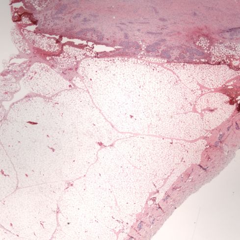
Figure 1: Prominent subcutaneous adipose tissue adjacent to the breast bud. (2x H&E)

Enter a caption
Figure 2: The inner third of the myocardium shows loss of basophilia in the myocyte nuclei, but nuclear basophilia is present in the outer third of myocardium. The dark granules are formalin hemoglobin pigment. (40x H&E)

Enter a caption
Figure 3: The liver shows partial loss of hepatocyte nuclear basophilia. The center shows a cluster of synchronous early erythroblastic cells consistent with increased erythroblastosis. (20x H&E)

Enter a caption
Figure 4: The lung demonstrates aspiration of more squames and debris than usual and there is a yellow cast to the cells. There were no dilated or plugged airways with meconium, but the findings are consistent with some meconium aspiration. (20x H&E)

Enter a caption
Figure 5: The arrows point to karyorhectic neurons in Sommer sector of the hippocampus. (40x H&E)

Normalized organ to brain weight ratios

Leave a comment