Archive | Uncategorized RSS feed for this section

Looking at fetal blood in the placenta

13 Jan

I decided to restart this blog, in part to find a way to post the material that was on my now defunct web site, but also because it seems every week if not every day there is some interesting problem or conundrum in obstetrical pathology that would be interesting to share with other others with an interest in obstetrical disease. That is the blog part. The more formal material will be in the pages and starts with a few pages of historical introduction to the discovery of an association of congenital birth injury with obstetrical events.

Placental sections provide an opportunity to look at fetal blood, admittedly inferior to a blood smear. I recent interesting case in point was the placenta from an almost term infant with a prenatal diagnosis of IUGR and of hepatomegaly. The villi appeared mature, but not enough to account for the growth restriction. The villi in the center of the placentone (spiral artery flow pattern) were slightly edematous. Perhaps, that is why I looked a little more closely at the fetal blood, which appeared hypercellular. This is often the low power observation that prompts a closer look for increased nuclear red cells. In this case, there was a more or less monomorphic mononuclear cell accumulation. This observation prompted bringing up the infant’s medical record with the thought that the cells might be the result of trisomy 21. There was no indication of Down syndrome, but the total white cell count was 229,000 with 45% blasts pre therapy.  The diagnosis was congenital acute B cell leukemia.

I at first was elated at having made an observation that correlated with the infant findings, but then was deflated that my diagnosis had not really added any useful information. Still, looking at fetal blood on the placental sections can add information and at the least confirm that the abnormal blood picture was congenital.

I have attached some microphotographs of the above case and other examples of fetal blood lesions in the placenta.


11275

This low power image shows the increased nucleated blood cells in the stem vessel.

11276

The fetal blood at first looked like increased nucleated red cells, but the cytoplasm was clear not red, and the overall impression was a monomorphic mononuclear population.


A case of familial congenital leukemia:


11289

Medium power showing leukemic cells in vessels but also in the villous stroma.

11287

High power showing the leukemic blasts.

A placenta from an infant with karyotype 48XXY+21

Blog 3

This placenta had scattered possible blasts and an abnormal blood picture but my notes are missing.

Parvo virus inclusion on H&E stain in an immuno-chemically proven case of infection.


parvo

The eosinophilic inclusions of parvo virus in the nucleated red cells.

Large red cells in triploidy

11293

The red cells are consistently larger in the fetal vessels compared to the maternal cells in the intervillous space in this 20 week fetus with dysgenic triploidy.

triploid coulter

On this Coulter strip, the red cell population on the left from the PUBS shows the large cells. The comparison on the right is after a neonatal red cell transfusion which show the normal peak and the residual triploid population.

Acanthocytosis in an infant with absent liver apoproteins

10098

The acanthocytes sometimes appear as knobs or as darker small spheres.

10102

Slightly less crowded together acanthocytes.

More on the adrenal, and three hypotheses

22 Feb

Because of the previous blog, a colleague Dr. Jeff Goldstein kindly shared a case of a 30 hour old infant with profound involution of the fetal zone of the cortex and prominent lipid in the permanent cortex, who died of severe congenital pulmonary malformation. The thymus did not show accelerated involution.

How is the involution of the fetal zone cortex controlled after birth? The biochemical evidence is that it ceases to function almost immediately although it takes days to weeks to demonstrate histologic involution. Some older papers suggested that HCG was trophic for fetal zone cortex, but not sufficient without ACTH to produce growth of this zone. Thus withdrawal of HCG with birth could be the signal for the involution of the fetal zone. If so there should be a correlation with Leydig cell involution which is HCG dependent (a luteinizing hormone analog). I searched my database for the lowest adrenal to brain ratio cases, and in the most extreme case, indeed there were no Leydig cells and the fetal zone had accelerated involution. This was a growth retarded 36 week gestation triplet infant without a clear cause of death at 30 minutes of life. I have no idea why this effect occurred, but these cases demonstrate that there are still mysteries to be solved.

Early evidence of involution of the fetal zone 40x

Early evidence of involution of the fetal zone 40x

Involution of most Leydig cells. 40x

Involution of most Leydig cells. 40x

However my time is finite and think I will shelve the attempt to write a book. Many placenta chapters are on my other website www.pediatricperinatalpathology.com. I also have chapters on infarction, subchorionic cysts and subchorionic thrombo-hematomas not posted.  I just never found a satisfactory way to post the extensive images that I wanted to use. I think the most valuable “chapters” were on maternal floor infarctions and amnion bands because I think I was able to synthesize some material well.

Instead of the book, I have three ideas that I want to concentrate on.

The first one is that the placenta is not only an obstetrical specimen but potentially a pediatric one.  This idea gained some support from the Barker hypothesis on the importance of intrauterine events on subsequent adult disease. I applied this idea to test two hypotheses to a large number of unselected placentas. The first sub-hypothesis that moderate to severe lymphohistiocytic villitis would correlate with childhood autoimmune disease was a complete negative. The second sub-hypothesis was that clinically silent old marginal separations with overlying large infarctions would predict neurologic deficits. The analysis has proved complicated and is still being pursued. However, to show clinical value for the placental examination in long term pediatric care is beyond my resources, and may not be true.

The second idea is that research needs to be directed at the natural causes of fetal asphyxia. Researchers have defined the nuances of controlled fetal asphyxia, but not really the natural mechanism. An important sub-hypothesis is that wrapping of the cord around the infant makes the effective cord length the distance from the placental insertion to the start of the wrap. This can create a short cord subject to torsion or compression from the wrapping that will compromise umbilical blood flow. I have seen more than once the scenario of a fetus with progressively worrisome fetal heart rate tracing who finally develops bradycardia and is rapidly delivered only to have hypoxic ischemic encephalopathy. The obstetrician notes a tight nuchal cord. It does not take much imagination to see that as labor progressed, this short cord by compression and torsion, was limiting umbilical blood flow, and likely at the end stopped all flow. This is a fairly good approximation to the primate model of Dr. Ron Myers of partial asphyxia followed by complete asphyxia as the mechanism that produces cerebral edema and severe neurologic injury. Unlike simple acute asphyxia there may be no window to rescue once bradycardia occurs. If the short free distance of the cord was known earlier, either elective Cesarean delivery or an intrauterine snare to unwrap the cord might have prevented disaster.  Unfortunately, I haven’t found an obstetrician willing to investigate the cause of fetal distress as soon as it becomes detectable.

The third idea is that we need to know the actual initiating mechanism that leads to preterm labor in order to prevent chorioamnionitis or ruptured membranes. I have discussed some of this in my blog.  I am working on a device to detect the earliest change of labor as a needed first step. This idea runs counter to those who blame chorioamnionitis as the cause of preterm labor. I think chorioamnionitis is part of the mechanism. I suspect the real root of preterm labor is in mechanisms developed to abort pregnancies when the maternal investment in the fetus was going to compromise the eventual survival of one offspring. This evolutionary approach is clearly presented in the book, “ Mother Nature: Maternal Instincts and How They Shape the Human Species”, by Sarah Hrdy.

I wish I was in an institution where research in these areas was fostered, and a pathologist’s input was welcome. There may be no such place, so I will continue to try to create one.

Stillborn autopsy case #2

21 Jan

This stillborn male infant was delivered by precipitous vaginal delivery at 33 weeks of gestation to his 30 year old, G3, Term 2, Living 2, Afro-American mother. Birth weight was 1,630 g. The mother’s prenatal care was unremarkable. She is blood type O+. The discharge summary diagnosed preeclampsia and dehydration. There was no Kleihaurer Bedtke test to detect fetal maternal hemorrhage, but a differential blood count on the mother after delivery had zero nucleated red cells.
The placental examination demonstrated a marked erythroblastosis (Fig 1). There was mild hydrops. There were no intranuclear inclusions typical of Parvo virus in the nucleated red blood cells, and an immunostain for Parvo virus was negative.

CA12-52_20xplac

The external examination demonstrated a hydropic male infant with prolonged retention based on extensive desquamation, and collapsed skull bones. However, the umbilical cord and abdomen were a pale pink, rather than the expected deep red (Fig 2). The gestational age exam demonstrated plentiful lanugo, 3-4 mm flat areola, descended testes, and slow recoil of the pinna. There were no plantar creases and no visible vessels, but these were not accurate parameters of gestation as the former appeared due to hydrops and desquamation and the latter due to the pallor. By cheating on the physical Ballard score, and giving 1 for both skin and plantar creases, the gestation is between 29 and 30 weeks, and giving 2 both for skin and plantar creases yields 32 weeks. The infant weighed 1,420 g at autopsy with loss of subcutaneous fluid weight into the blankets which likely accounts for the difference from the birth weight. The autopsy weight is appropriate for this range of gestations. There were no malformations.

Fig 2 Infant showing extensive desquamation and pale red color of abdomen and cord

CA52 (1)

The internal examination demonstrated only small pleural effusions. The thymus appeared small and the spleen large. The heart was normal but the ductus arteriosus appeared relatively narrow but still more than 50% of the descending aortic diameter. The brain was very soft but not liquid and the convolution development was appropriate for 30-32 weeks of gestation. There were no malformations of any organ.

The weights are listed in table 1. Since the relative weights are usually most informative, I have calculated the organ to brain weight ratios and then subtracted these from the expected ratios, then divided that difference by the expected ratio[1]. The graph of this calculation makes interpretation of the weights easier (Fig 3). The kidney and spleen are clearly larger than they should be. The heavier adrenals and lighter thymus are consistent with stress, adrenal lipid accumulation and thymic loss of cortical lymphocytes, but not very marked. The lungs are not small, as would be expected had there been a persistent pleural effusion and this result correlates with the small effusion and normal lung size seen grossly.

Table 1
Autopsy
weight Brain Lungs Heart Kidneys Adrenals Liver Spleen Thymus
1420 270 52.8 14.1 33.0 7.8 113.3 24.1 4.2

ca53tabwt tablele

The microscopic evaluation of intrauterine postmortem retention based on the publication of Genest and colleagues was at least one week with complete loss of basophilia in the liver, adrenal and ileocecal sections, and loss of tracheal cartilage basophilia[2]. The external features did not appear to be extreme and the kidney was microscopically relatively intact, suggesting that in the more than 1 week to less than 4 week Genest interval of dating, this infant was closer to the one week.
The thymus showed a modest loss of cortex microscopically and the adrenal could not be easily evaluated, and there was no frozen sample for fat staining, but the findings were not those of prolonged fetal stress as would have been expected in Rh alloimmunization. The kidney interstitium demonstrated plentiful hematopoiesis which appeared to account for the increase in weight. As is typical in the kidney, the hematopoietic response included myelopoiesis (Fig 4). The enlarged spleen was due to a cellular infiltration not just congestion, but the autolysis compromised interpretation. An iron stain highlighted a diffuse distribution of hemosiderin macrophages that were no very evident on H&E stain (Fig 5).

Kidney showing hematopoiesis around a vessel, H&E 20x

[caption id="attachment_58" align="alignnone" width="300"]Spleen, left H&E 20x, right iron stain 40s showing pale hemosiderin macrophages. Spleen, left H&E 20x, right iron stain 40s showing pale hemosiderin macrophages.

Kidney showing hematopoiesis around a vessel, H&E 20x[/caption]

The placenta had a mild maternal chorioamnionitis and despite an absence of neutrophils in the fetal lung or stomach, postmortem lung cultures grew scant Corynebacterium species. This inflammation was likely postmortem from the fetal perspective, but may have facilitated the onset of labor.

The underlying diagnosis in this infant was a relatively acute onset of hydrops related to a drop in hematocrit and likely high output heart failure, based on the marked erythroblastosis in the placenta, and the pale edematous fetus.
There are four main differential considerations of the cause.

1. Hemorrhage: There was no internal hemorrhage. There was no evidence of hemosiderin or phagocytized red blood in the membranes, nor any cause of hemorrhage seen grossly in the placenta, e.g. velamentous vessels, or chorionic vessel aneurysm, making hemorrhage external into the amnion space or vagina unlikely. There was no maternal blood test for fetal blood in the mother. Since her blood count had no nucleated red cells, it is unlikely that there was any recent hemorrhage since the fetus had approximately 25% of his red cells nucleated. Against fetal maternal hemorrhage is the intensity of the erythroblastosis, the splenomegaly, and most strongly the hemosiderin in the spleen.
2. Infection: Parvo virus is the most likely cause of relatively acute anemia, but there was no evidence for that infection, and there should have been if it was present. There was no evidence of other fetal infection.
3. Hemoglobinopathy: I have no personal experience with Barth’s hemoglobin, but I would not expect it to have evidence of the short duration of hydrops seen in this infant. The patient is Afro-American and there was no family history of anemia in the chart, but I can not completely exclude the diagnosis.
4. Hemolysis: The hemosiderin in the enlarged spleen in good evidence of the phagocytosis of injured red cells. The mother is Rh negative, and has had two living children. The mother has type O blood and a recent exposure to type A or B might have produced IgG hemolytic antibodies to a major blood type, or she may have antibodies to a non-Rh blood antigen. The autopsy report recommended testing mother for hemolytic antibodies. The fetal blood type was not obtained, but in theory some red blood cell antigens may be detectable on the placental tissue sections. The fetal cells were extremely autolytic, but the maternal blood flow of the placenta maintains some of the red cells despite karyorhexsis.

Tentatively, the final diagnosis is hemolytic disease of unknown cause with a short duration of fetal hydrops and anemia. I would appreciate any comments or help with this case.

1. Fujikura, T. and L. Froelich, Organ-weight/brain -weight ratios as a parameter of prenatal growth: A balanced growth theory of visceras. Am J Obstet Gynecol, 1972. 112: p. 896-902.
2. Genest, D.R., M.A. Williams, and M.F. Greene, Estimating the time of death in stillborn fetuses: I. Histologic evaluation of fetal organs; an autopsy study of 150 stillborns. Obstet Gynecol, 1992. 80: p. 575-84.

Why do primates have a fetal adrenal?

16 Jan

The adrenal gland in the human fetus is proportionately much larger compared to the kidney than it is in the child or adult[1]. This increase in size is due to the fetal adrenal, a histologically distinct inner region that compromises over 80% of the gland and produces 16-OH-Dihydroepiandrosterone sulfate (DHEAS), the necessary precursor for the placental production of estrogen which is secreted into the maternal circulation. This fetal adrenal is a feature of primates but not of other mammals.

In the sheep, Liggins and colleagues discovered that the placenta has the enzyme, CYP17, which is absent in the primate placenta, and is necessary to convert cholesterol via pregnenolone into estrogen[2]. When the lamb hypothalamic axis matures, it stimulates cortisol production in the adrenal via ACTH.  Cortisol induces organ maturation in preparation for birth, and up-regulates CYP17 resulting in increased maternal estrogen.  The increasing ratio of estrogen to progesterone is the signal for myometrial cells to alter their phenotype via gap junctions, receptors and calcium channel modifications to produce labor.  Why did primates abandon this simple mechanism of estrogen production for the more circuitous fetal adrenal function?

The speculative answer is that primates had large brains and needed to be delivered prior to full maturation of the brain. If not, the large infant head would become entrapped in the pelvis. Survival of the big brained primates would require the co-evolution of a way to deliver this big brain. The fetus needed to be delivered before the hypothalamic axis was fully mature, but cortisol would still be needed to mature other organs especially the immature lungs.

To deliver the big brained infant prematurely, cortisol production needed to be uncoupled from brain maturation. The primate may accomplish this uncoupling via corticotrophin releasing factor (CRF) produced in the placenta rather than from the mature hypothalamus[3].  This mechanism may have evolved originally to abort compromised pregnancies. For example if the placenta became relatively hypoxic, sending a placental generated CRF signal to the pituitary would increase ACTH and hence cortisol. This would initiate labor and result in the premature delivery of the fetus with the added benefit of a boost to its lung maturation. The spontaneous abortion of a compromised fetus might improve the mothers overall reproductive success. Even If we accept the speculation that the initiation of labor in the primate hijacked a local placental mechanism of CRF to initiate labor several weeks earlier than brain maturation would have, we still need a rationale for abandoning the placental production of estrogen. Placental CRF could still have initiated the cortisol dependent CYP17 mechanism of labor as in the sheep.

Speculatively, the rise in cortisol from placental CRF may have needed more time to act than in a more mature infant and the placental production of estrogen was simply too rapid a response.  The function of the fetal adrenal may have been to delay the estrogen production compared to the placenta. The response of the fetal adrenal to ACTH is production of DHEAS but there is a damping inhibition of estrogen on that response. The problem with this theory is that in the primate, labor can occur without fetal adrenal function.

Stillborn infants still deliver from spontaneous labor.  Infants with anencephaly do not develop a large fetal adrenal but still deliver spontaneously.  At most, anencephalic infants have a pregnancy extended by 2 weeks, and this is as likely related to delayed ratio of oxygen use to placental function,  as due to lower estrogen [4].  Adrenalectomy in fetal monkeys does not significantly prolong labor[5]. Fetal sulfatase deficiency which blocks the maternal elevation of estrogen  production is not associated with prolonged gestation[6]. However there is other evidence that supports a role for the fetal adrenal in initiating labor.

The evidence for a role of the fetal adrenal in labor initiation in humans and monkeys starts with the observation that maternal serum estrogen and cortisol increase before and during labor. This does not prove causation, but requires an explanation.  In monkeys, fetectomy prolongs labor which contradicts the study on adrenalectomy [7]. In humans, some intrauterine deaths do not lead to labor as evidenced by the existence of defibrination syndrome that is prevented by inducing labor that has become too delayed. Infants with sulfatase deficiency may require more Cesarean sections for failed labor. Thus, there may be a role for the fetal adrenal but it is not absolutely required to produce labor.

The most clear demonstration of a key role for fetal androgen as a precursor to estrogen in labor is that the direct intravenous infusion of androstenedione in monkeys produces early onset of normal labor compared to controls[8, 9]. (The authors cite a reference in a monograph that infusion of DHEAS does not increase maternal estrogen in monkeys, but no explanation is given.) Androstenedione like DHEA can be converted by aromatase in the placental syncytiotrophoblast to maternal estrogens. When these studies were repeated with an aromatase inhibitor to prevent conversion of the androgen to estrogen, as expected maternal estrogen did not increase, nor did premature labor ensue. However, when monkeys were infused with estradiol to produce a rise in estrogen comparable to that in labor, only myometrial contractions occurred but no cervical change and no preterm delivery. In none of the infusion experiments did progesterone decrease. The authors conclude that there must be paracrine effects at the local site of production[10]. An alternative hypothesis is that the conversions from androgens to estrogens regulate other processes that simply are not being measured in the experiments.  The infusion experiments suggest that the fetal adrenal – estrogen pathway can initiate term labor.

In favor of the importance of the fetal adrenal is the investment of fetal resources to create such a large metabolically active organ.  The CRF to ACTH to DHEAS to estrogen pathway plausibly can initiate labor, but the function of the fetal adrenal placental system is more complex than just the production of estrogen, as the androgen infusion experiments indicate. For example some DHEA goes to the fetal liver to produce estriol, not just to the placenta to produce maternal estrogen. As many others have observed, the existence of redundancy would be expected in so important a survival function as initiating labor. Fetal adrenal function may be a primary but not an exclusive initiator of term labor.

The progesterone receptor blocking drug RU 486 stimulates uterine contraction but not cervical change[11]. Infused estrogen can produce myometrial contractions, but does not dilate the cervix. Iatrogenic prostaglandins do dilate the cervix and initiate labor. CRF up-regulates membrane cyclooxygenase and this could be the link to successful labor using both estrogen and prostaglandin, as well as the mechanism to produce labor in the absence of fetal adrenal function.

If placental CRF is the main signal initiating term labor in humans as opposed to hypothalamic maturation, then what determines the timing of CRF secretion? If CRF can stimulate cyclooxygenase, where does this occur and how is it amplified? As noted in the chorioamnionitis discussion, it is possible that chorioamnionitis is a method for the uterus to amplify the cervical signal with increased inflammatory mediators induced by usually benign vaginal flora. These are important questions, particularly in relation to premature labor that need to be considered in future blogs.

Bottom line: The question of why do we have a fetal adrenal is ultimately a question of its fitness for survival, and I have speculated on how and why it may have come about. There is some evidence that its production of DHEA-S with conversion to estrogen in the placenta is stimulated by placental CRF as a stimulus for the initiation of labor.


1.            Langlois, D., J.Y. Li, and J.M. Saez, Development and function of the human fetal adrenal cortex. J Pediatr Endocrinol Metab, 2002. 15 Suppl 5: p. 1311-22.

2.            Liggins, G.C., et al., The mechanism of initiation of parturition in the ewe. Recent Prog Horm Res, 1973. 29: p. 111-59.

3.            Giannoulias, D., et al., Localization of prostaglandin H synthase, prostaglandin dehydrogenase, corticotropin releasing hormone and glucocorticoid receptor in rhesus monkey fetal membranes with labor and in the presence of infection. Placenta, 2005. 26(4): p. 289-97.

4.            Milic, A.B. and K. Adamsons, The relationship between anencephaly and prolonged pregnancy. J Obstet Gynaecol Br Commonw, 1969. 76(2): p. 102-11.

5.            Mueller-Heubach, E., R.E. Myers, and K. Adamsons, Effects of adrenalectomy on pregnancy length in the rhesus monkey. Am J Obstet Gynecol, 1972. 112(2): p. 221-6.

6.            Bedin, M., et al., Incidence of placental sulfatase deficiency on the mode of termination of pregnancy. Gynecol Obstet Invest, 1987. 24(2): p. 86-91.

7.            Nathanielsz, P.W., J.P. Figueroa, and M.B. Honnebier, In the rhesus monkey placental retention after fetectomy at 121 to 130 days’ gestation outlasts the normal duration of pregnancy. Am J Obstet Gynecol, 1992. 166(5): p. 1529-35.

8.            Mecenas, C.A., et al., Production of premature delivery in pregnant rhesus monkeys by androstenedione infusion. Nat Med, 1996. 2(4): p. 443-8.

9.            Figueroa, J.P., et al., Effect of a 48-hour intravenous delta 4-androstenedione infusion on the pregnant rhesus monkey in the last third of gestation: changes in maternal plasma estradiol concentrations and myometrial contractility. Am J Obstet Gynecol, 1989. 161(2): p. 481-6.

10.          Nathanielsz, P.W., et al., Local paracrine effects of estradiol are central to parturition in the rhesus monkey. Nat Med, 1998. 4(4): p. 456-9.

11.          Haluska, G.J., et al., Temporal changes in uterine activity and prostaglandin response to RU486 in rhesus macaques in late gestation. Am J Obstet Gynecol, 1987. 157(6): p. 1487-95.


Chorangiosis?

11 Dec

Dr. Altshuler’s brief often cited paper in 1984 gave a precise definition of chorangiosis [1]. Despite the definition’s fame as a series of tens, the definition is somewhat arbitrary* and the clinical correlations nebulous. Chorangiosis has become an accepted name for a pathological observation that in some form certainly exists. An earlier paper by Caldwell et. al. attributes what seem to be the first English use of the term, chorangiosis, to a personal communication with Dr. Shirley Driscoll[2]. I may be an outlier, but I am still uncertain what chorangiosis implies as a diagnosis.

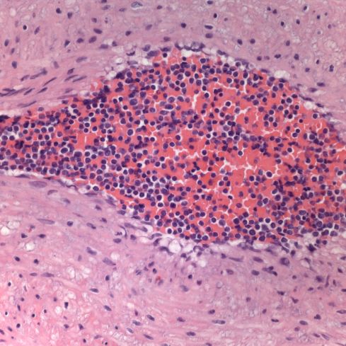
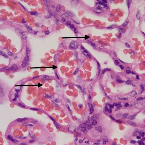
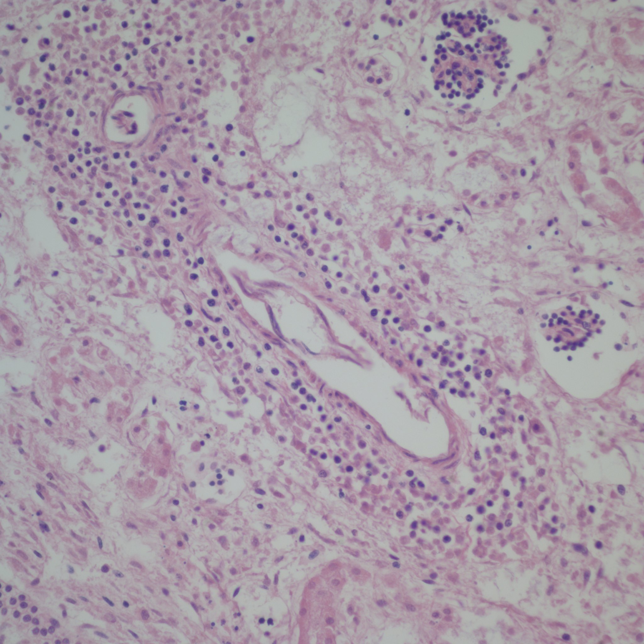
First I have trouble distinguishing hypervascularity in the villi from congestion which likely increases the apparent number of capillaries seen in microscopic cross section of a villus due to folding of engorged capillaries. Dr. Altshuler’s paper, warned of possible confusion between the two, and provided an illustration. Looking at his comparison photomicrograph in the paper I count at least 6 villi with more than 10 vascular profiles in the congested villi example. Given a photograph from a different area, that case might have qualified as chorangiosis. Many of the published papers illustrating chorangiosis in fact show villi with very dilated capillaries that do not give me confidence in the diagnosis[3-5]. To quote from a case review of chorangiosis: “Therefore, rigid diagnostic criteria were established by Altshuler (1984) for diagnosis of CH. These criteria, though difficult to apply, aim at differentiating CH from congestion(vessels numerically normal) and tissue ischemia (shrinkage of villi)…[6]”. The underline is mine. The acute capillary dilatation and probably increased blood flow over a retroplacental hemorrhage appears to increase the number of capillaries (fig) 1. This may explain the reported association of chorangiosis and abruption[7]. Perhaps congested villi with apparent hypervascularity should tentatively be a separate entity.

Dilated villous capillaries above a retroplacental hematoma

Dilated villous capillaries above a retroplacental hematoma


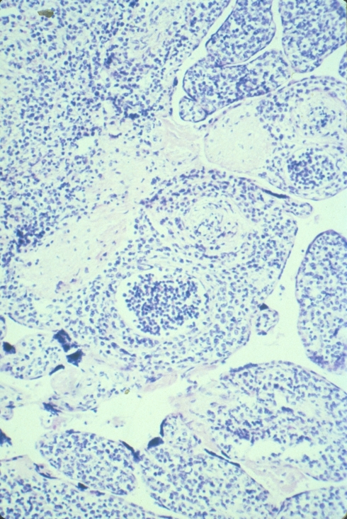
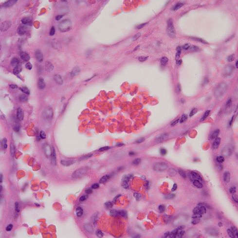
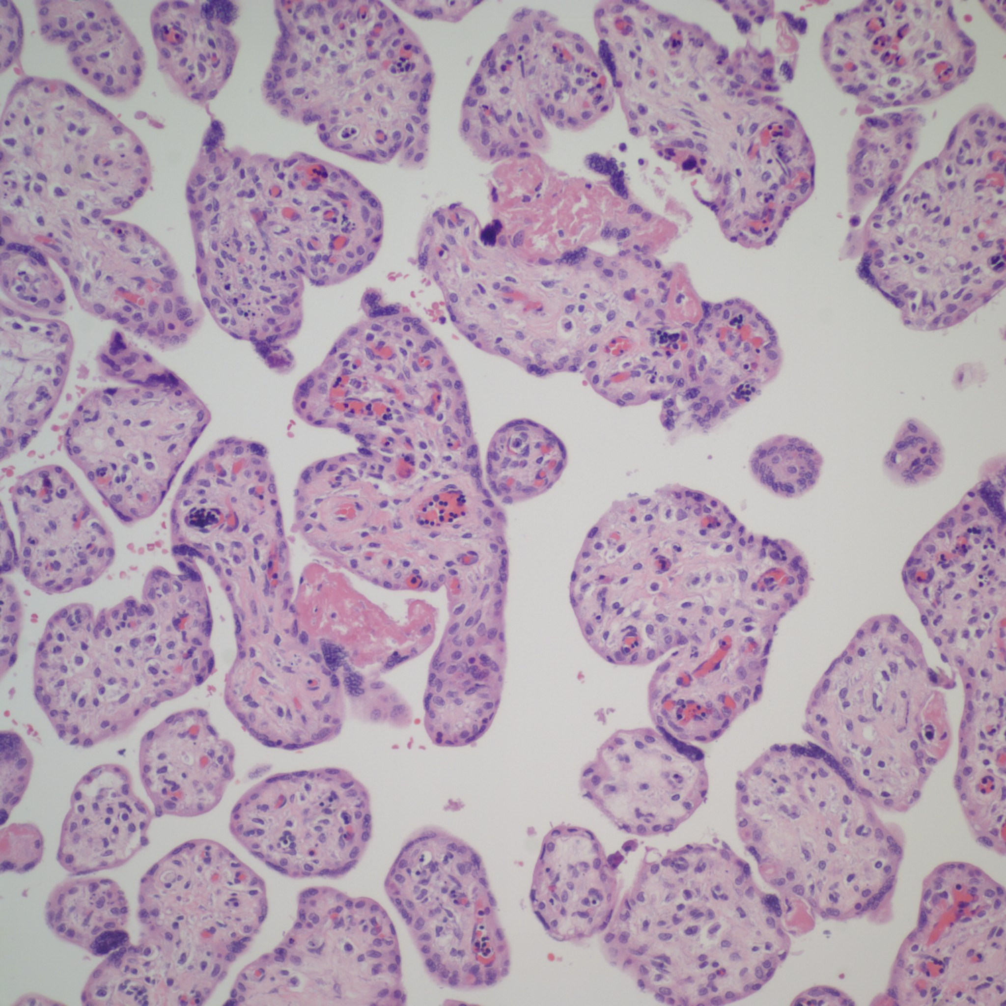
Distinguishing chorangiosis with its large terminal villi from persistent or increased intermediate villi is also not simple. I suspect that this confusion may be responsible for the association of maternal diabetes and chorangiosis. Dr. Ogino and Redline’s paper specifically mentions that the association is also with placentomegaly and delayed villous maturation. This problem can be seen clearly in looking at very early gestation placentas. (fig 2)

Placental villi from an aborted Potter syndrome fetus at 23 weeks gestation

Placental villi from an aborted Potter syndrome fetus at 23 weeks gestation

Another problem is to distinguish chorangiosis from diffuse chorangioma (chorangiomatosis), which was amply clarified in the same Orgino and Redline paper. Yet this confusion persists, at least in the titles, in some publications [8, 9].


            Chorangiosis currently does not imply a distinct pathogenesis. The associations in Dr. Altshuler’s paper were with very broad clinical categories, things like neonatal death 39% positive, anomalies 27% positive, and Cesarean section (32%). The significance of the high percentages must be tempered by the fact that the placentas were examined for obstetrical indications. Dr. Altshuler states that  “My hospital rarely encounters it in normal pregnancies.” This is less than valid statistical analysis.

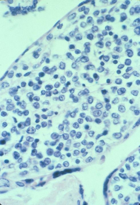
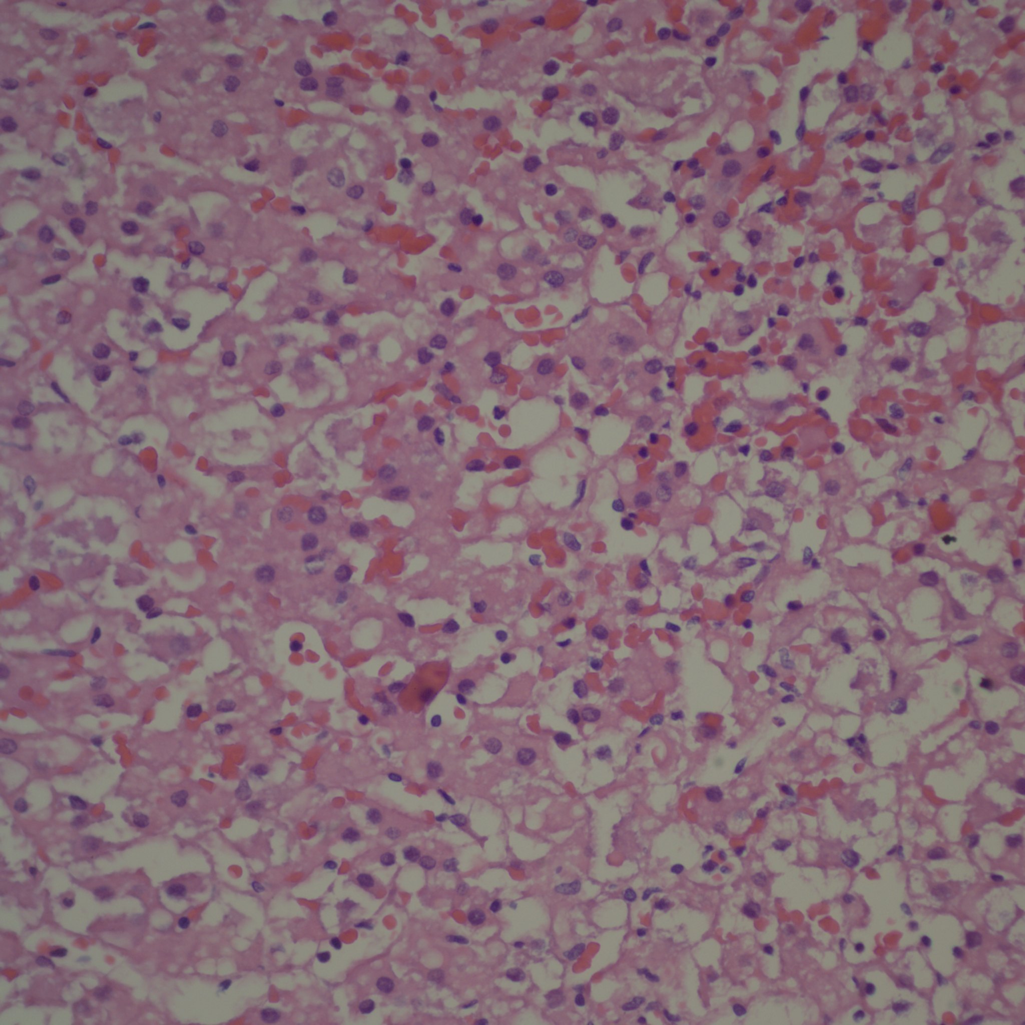
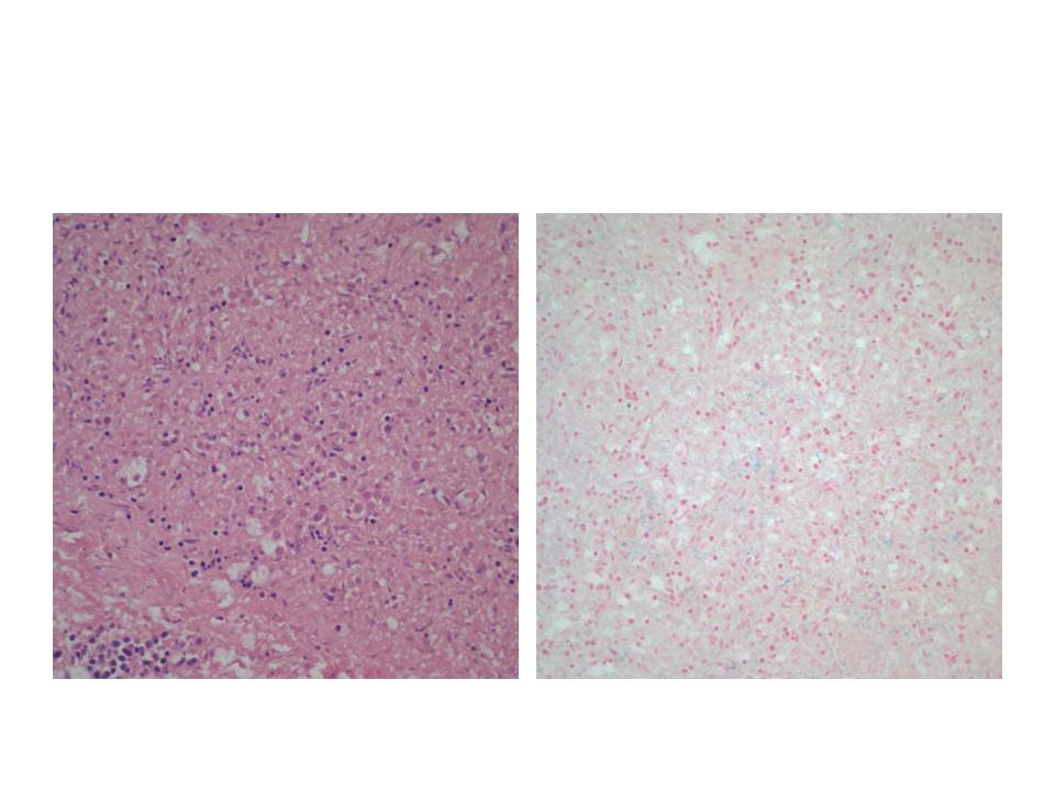
Altshuler’s paper found that 27% of cases had villitis of unknown etiology. This is certainly a finding that routine placental examination appears to support (fig 3). In practice, if I see a focus of larger, hypervascular villi, this is a clue to look for villitis and avascular villi. I rationalize this hypervascularity as due to increased blood flow in a segment of a stem villus in which other segments have capillary occlusion. The villi with shunted blood flow adapt over time by producing more capillaries. A similar adaption to high flow can be seen in diffuse hypervascularity of villi in the recipient twin in twin-to-twin-transfusion in which the pregnancy was prolonged with serial amniocentesis[10].

Lympho-histiocytic villitis and hypervascular villi

Lympho-histiocytic villitis and hypervascular villi

In a previous blog I have discussed the papers by Mana Parast and colleagues associating umbilical cord occlusion with fetal vascular thrombi and avascular villi [11-13]. If focal chorangiosis accompanies such avascular villi this might explain the association with Cesarean section if they were performed for fetal distress. Some have found cases of chorangiosis with umbilical cord abnormalities, for example tight nuchal cord[14]. Such a relationship could tie avascular villi, chorangiosis and poor fetal outcomes together, but it would need to be tested. Even so, a reasonable first draft might separate focal hypervascular villi associated with villitis or avascular villi as a separate entity from a diffuse form.

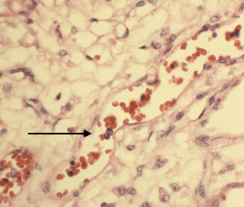
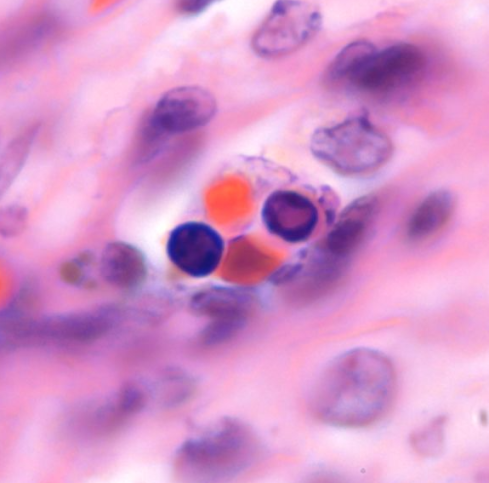
While some have suggested chronic hypoxia as a cause of chorangiosis, I think that is unlikely for two reasons [4, 5, 15-17]. First I don’t see hypervascular villi with chronic utero-placental ischemia even in stillborn infants, many of whom likely had become hypoxic.  Second, increasing the capillaries does not necessarily increase oxygen transport to the fetus. See Mayhew and colleagues discussion of the relative role of parameters of oxygen transfer in the placenta[18]. Keeping most parameters within physiological limits, the only important variable is the thickness of the barrier between the maternal and fetal circulation. Another possible mechanism of chorangiosis is a varicose vein of the villi mechanism in which back venous pressure is the cause. I think this is unlikely since the usual effect of elevated umbilical venous pressure is hydrops. A third theory is a genetic or acquired mismatch in growth factors, e.g. witness the extreme capillary proliferation in mesenchymal dysplasia of the placenta of which one cause is the imprinting error in Beckwith-Wiedemann syndrome. There is a case report of chorangiosis associated with elevated maternal HCG late in pregnancy[19], which may be a clue to a type of mechanism. The following figure 4 shows the abrupt onset of capillary proliferation from a stem villus in a placenta submitted for “probably Down Syndrome” that might have a genetic basis.

Normal stem villus with attached branch showing capillary proliferation

Normal stem villus with attached branch showing capillary proliferation

The very concept of chorangiosis suggests a mismatch between capillary and villous growth. Growth factors favoring endothelial proliferation are likely involved. New branching would not be necessary since stuffing a longer capillary in the villus is likely to curl up and increase capillaries per cross section. However, there could be differences in the three dimensional anatomy of the blood vessel growth that would be difficult to discern in a microscope section, and such differences might reflect some aspects of pathogenesis.

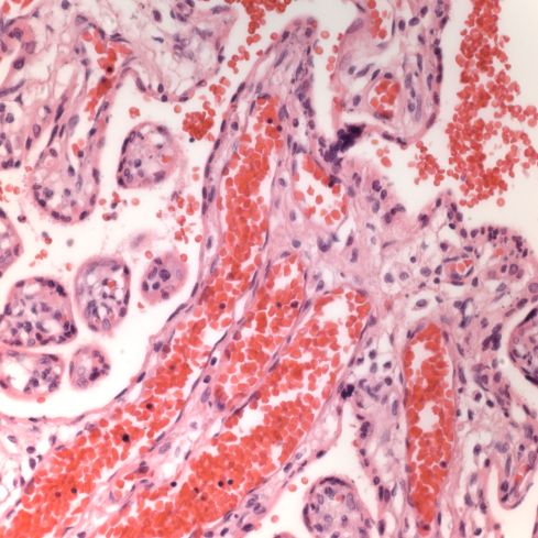
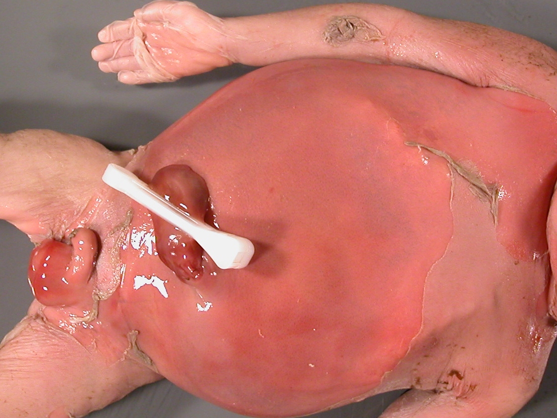
I recently tried to look at a placenta with diffuse villous hypervascularity under the dissecting scope( fig 5,6,7). I wanted to compare my case to the classical SEM corrosion casts of villous capillaries but without the work[20]. It was a total disappointment. After fixation the blood was not visible in the capillary vessels. This case is interesting because the placenta was submitted to pathology by an obstetrician who submits all placentas even without a specific obstetrical problem. The infant was an AGA 39 week vaginal delivery with Apgars of 8 and 9 at 1 and 5 minutes without recorded complications. However the placenta weighed 790 g for a very abnormal fetal placenta weight ratio of 4.3, reflecting the abnormal villi.

Diffuse increased vascularity

Diffuse increased vascularity

Diffuse increased vascularity

Diffuse increased vascularity

Dissecting scope of same hyerpvascular villi

Dissecting scope of same hyerpvascular villi

I diagnosed the above case as a mature placenta with hypervascular villi. I commented that the significance of the lesion is unknown.

 Bottom Line In general I think the criteria of Altshuler are too inclusive and prone to overlap normal cases especially with villous congestion, so I do not make a diagnosis “chorangiosis”.I propose a tenative reclassification of “Altshuler’s lesion”1. Focal hypervascular villi associated with villitis2. Focal hypervascular villi associated with avascular villi3. Hypervascular villi with marked capillary dilatation4. Hypervascular villi associated with delayed villous maturation5. Diffuse hypervascular villi without other features. At this point I think the diagnosis should depend on a clear separation from any normal distribution of villous capillaries, but I don’t have a simple forumula.              I look forward to any disagreements, suggestions or examples.


Bob Bendon 

 *“Chorangiosis was diagnosed when inspection with a 10x objective showed tem villi, each with ten or more vascular channels in ten of more non-infarcted and non-ischemic zones of at least three different placental areas.”

References:

1.         Altshuler, G., Chorangiosis. An important placental sign of neonatal morbidity and mortality. Arch Pathol Lab Med, 1984. 108(1): p. 71-4.

2.         Caldwell, C., et al., Chorangiosis of the placenta with persistent transitional circulation. Am J Obstet Gynecol, 1977. 127(4): p. 435-6.

3.         De La Ossa, M.M., B. Cabello-Inchausti, and M.J. Robinson, Placental chorangiosis. Arch Pathol Lab Med, 2001. 125(9): p. 1258.

4.         Soma, H., Y. Watanabe, and T. Hata, Chorangiosis and chorangioma in three cohorts of placentas from Nepal, Tibet, and Japan. Reprod Fertil Dev, 1995. 7(6): p. 1533-8.

5.         Akbulut, M., et al., Chorangiosis: the potential role of smoking and air pollution. Pathol Res Pract, 2009. 205(2): p. 75-81.

6.         Gupta, R., et al., Clinico-pathological profile of 12 cases of chorangiosis. Arch Gynecol Obstet, 2006. 274(1): p. 50-3.

7.         Staribratova, D. and N. Milchev, [Placental chorangiosis associated with abruption and hypoxia]. Akush Ginekol (Sofiia), 2009. 48(5): p. 44-6.

8.         Caldarella, A., A.M. Buccoliero, and G.L. Taddei, Chorangiosis: report of three cases and review of the literature. Pathol Res Pract, 2003. 199(12): p. 847-50.

9.         Schmitz, T., et al., Severe transient cardiac failure caused by placental chorangiosis. Neonatology, 2007. 91(4): p. 271-4.

10.       Pietrantoni, M., et al., Mortality conference: twin-to-twin transfusion [clinical conference]. J Pediatr, 1998. 132(6): p. 1071-6.

11.       Tantbirojn, P., et al., Gross abnormalities of the umbilical cord: related placental histology and clinical significance. Placenta, 2009. 30(12): p. 1083-8.

12.       Parast, M.M., C.P. Crum, and T.K. Boyd, Placental histologic criteria for umbilical blood flow restriction in unexplained stillbirth. Hum Pathol, 2008. 39(6): p. 948-53.

13.       Ryan, W.D., et al., Placental histologic criteria for diagnosis of cord accident: sensitivity and specificity. Pediatr Dev Pathol, 2012. 15(4): p. 275-80.

14.       Franciosi, R.A., Placental pathology casebook. Chorangiosis of the placenta increases the probability of perinatal mortality. J Perinatol, 1999. 19(5): p. 393-4.

15.       Schwartz, D.A., Chorangiosis and its precursors: underdiagnosed placental indicators of chronic fetal hypoxia. Obstet Gynecol Surv, 2001. 56(9): p. 523-5.

16.       Suzuki, K., et al., Chorangiosis and placental oxygenation. Congenit Anom (Kyoto), 2009. 49(2): p. 71-6.

17.       Barut, A., et al., Placental chorangiosis: the association with oxidative stress and angiogenesis. Gynecol Obstet Invest, 2012. 73(2): p. 141-51.

18.       Mayhew, T.M., M.R. Jackson, and J.D. Haas, Microscopical morphology of the human placenta and its effect on oxygen diffusion: a morphometric model. Placenta, 1986. 7: p. 121-131.

19.       Smith, A.S., et al., Placental chorangiosis associated with markedly elevated maternal chorionic gonadotropin. A case report. J Reprod Med, 2003. 48(10): p. 827-30.

20.       Habashi, S., G.J. Burton, and D.H. Steven, Morphological study of the fetal vasculature of the human term placenta: scanning electron microscopy of corrosion casts. Placenta, 1983. 4(1): p. 41-56.


Maternal Floor Infarction (MFI) demystified?

3 Dec

I was pleased to see that the paper on LCDH mutations and maternal floor infarction was finally published[1]. The material had been presented at the Philadelphia SPP meeting. This is a paper of courage and persistence in tracking down a long shot based on a single case report.

By now everyone agrees that maternal floor infarction is a misnomer at least for most of the cases so labeled. The pathology is a diffuse increase in peri/inter villous fibrinoid and usually with some intervillous thrombi. To understand the lesion, the conditions in which perivillous fibrinoid forms must also be understood. I think this understanding can be achieved following the advice of Yogi Berra “You can observe a lot just by watching”.

We see fibrinoid at the edges of old infarctions. The center of the infarction has lost all  blood flow and shows a collapsed intervillous space and complete coagulation of the villi, outside of which is a shell of perivillous fibrinoid with entrapped villous cores, and then villi adapting to low blood flow that are narrow with increased syncytial knots (yet another misnomer) and capillary syncytial membranes.  A similar pattern can be seen around old intervillous thrombi. There are stalactites of fibrinoid dripping from the subchorionic plate where they seem to follow the likely low flow areas from the fountain like spiral artery perfusion of the intervillous space. To me the common factor in all of these locations is an intermediate degree of hypoxia between coagulation necrosis of syncytium and syncytial adaptation.

The next relevant observation is at the edges of intervillous fibrinoid particularly in areas of massive perivillous fibrinoid deposition. The edges show apoptotic appearing condensation of the syncytial nuclei and apparent peeling off the syncytium.  The result is an exposed surface of the villous stroma with preservation of the subsyncytial Langhans cytotrophoblast . The process might be an overly exuberant expelling of synctytial knots. The end result of such exposure is cytotrophoblast proliferation and production of surrounding extracellular matrix and the layering of fibrin from the intervillous space. If isolated the villous surface is “re-epitheliazed”  by syncytium,  but in rapidly forming deposition the matrix and fibrin contact the surface of another denuded villus and the area of fibrinoid deposition enlarges.

Okay, so this is a lot of speculation based on some still H&E pictures, but it is does provide the basis for a consistent hypothesis about the formation of massive perivillous fibrinoid deposition. The theory is that anything that kills the synctytial surface of the villi but leaves the cytotrophoblast intact can produce massive fibrinoid deposition. Antibodies to anti-paternal antigens, or even autoantibodies, on the syncytial surface could result in syncytial death. Repetitive hypoxia that can propagate itself as I saw in a case of multiple sickle cell crises might produce the same lesion. In rats a similar lesion can occur with an oxidative poison. Which brings us back to the recent  paper.  I would like to think that the mutation in beta oxidation of lipid simply leaves the syncytium energy depleted. Normally it is the most intense consumer of oxidative metabolism of glucose in the body, presumably because of the transport needs of the fetus.

This would make such a neat theory, but there is a major wrinkle. The mutations in HADHA in the MFI cases were a strange mix of novel mutations. Is there a common thread among these mutations?  Beats me.  MFI is not, to my knowledge or experience, associated with acute fatty liver of pregnancy or HELLP syndrome. There is something unexplained going on here that begs for more analysis. I have offered to submit some blocks to Linda Ernst, and I know others have also volunteered.  The problem as I understood it some years ago was funding. Is there no kind soul out there who can undertake the possibly frustrating task of sequencing the HADHA gene from formalin fixed tissue blocks?

A prospective study of MFI measuring the metabolism of living villi in vitro with known could test the hypothesis.  I was involved in the paper by Mandsager showing that the diagnosis of MFI can be made prenatally, so there would be time to set up the experiment up[2].

References:

For a more complete literature review see our group’s website www.pediatricperinatalpathology.com, under placenta

1.            Griffin, A.C., et al., Mutations in long-chain 3-hydroxyacyl coenzyme a dehydrogenase are associated with placental maternal floor infarction/massive perivillous fibrin deposition. Pediatr Dev Pathol, 2012. 15(5): p. 368-74.

2.            Mandsager, N.T., et al., Maternal floor infarction of the placenta: prenatal diagnosis and clinical significance. Obstet Gynecol, 1994. 83(5 Pt 1): p. 750-4.

Machin comment on stillborn case posted on New Venue post, see there

30 Nov

Dr. Geoff Machin commented on the stillborn case, but somehow the comment attached to the new venue post. His argument is that cord torsion is very important and the vein is more vulnerable than the arteries to torsion. I don’t disagree. What I do have a problem with is the value of the cord coiling index. I think the pitch of acute torsion is more affected by the length of cord free to twist than any history of torsion embedded in the cord structure. But all of this needs some good experimental work which could be done. I will expand on this for Geoff in another post so that I can see where our differences and similarities really are. I also think that if stasis occurs because of venous occlusion, flow will slow everwhere in the system, and where thrombosis occurs may be more related then to vascular injury from turbulence induced stasis and from tissue hypoxia. Arterial emboli to the placenta can occur, we published a case of fetal aortic thrombus that appeared to be guilty, but it may not be an important mechanism. I really should paint the vessels grossly where it is easy to tell vein from artery, to identify them histologically and get a better handle on all this. More later.  Thanks Geoff for responding, and I hope others will read it and think about it.

Stillbirth case: umbilical cord “accident”

20 Nov

Case of stillbirth

This stillborn infant was delivered vaginally at 35 weeks five days gestation to a G2 P1 mother with a previous fetal demise. The female infant weighed 2,090 g. Mother smoked occasionally during pregnancy. Based on an ultrasound examination at 11 menstrual weeks of gestation, her expected delivery date was moved forward 2 weeks. She had fetal heart tones 2 weeks before an exam that had no fetal beat. She was delivered 24-48 hours later. Her prenatal care was otherwise unremarkable. There are no previous specimens in the pathology database for this patient.
Her placental examination showed a 390 g placenta with a pale area 5 cm in diameter that histologically showed hemorrhagic endovasculosis, but no thrombi in fetal vessels. The diagnosis was evidence of fetal vascular occlusion but no thrombi were seen in the sample. There was also mild erythroblastosis.
The gross examination of her autopsy demonstrated pleural petechiae, and small (10-20 ml) pleural and pericardial serous infusions. An unusual finding was subcutaneous petechial hemorrhages over the anterior chest. Microscopically, beside confirming the petechiae and using the criteria of Genest for histologic dating, nothing in the body was remarkable. The findings were consistent with 24-48 hours of postmortem retention.
The brain notably demonstrated very extensive karyorhexsis of nuclei in the basis pontis and in the subiculum.
A relook at the gross placenta failed to find the pale area and could find no tell-tale hemoglobin staining of surface vessels or palpably firm thrombi in those vessels.       However after several days of fixation in formalin, there were small suspicious pale areas. The placenta was resampled extensively including the insertion of the umbilical cord and the large surface vessel branches.
Microscopically, the pale areas were small areas of avascular villi (fig 1), but the volume of involved villi was small probably less than 1 percent of the placenta, and since these were fibrotic would have been likely days or more in duration. The real surprise was that almost all of the large vessels including the insertion showed non-occlusive mural thrombi sometimes with early organization(fig 2).


Fig 2: Mural thrombus in a surface vessel

Fig 1: Avascular villi on the right, slightly hypervascular villi on the left


So how does a pathologist interpret these findings to explain the fetal death?

First, I have argued elsewhere, the intrathoracic petechiae are evidence of fetal gasping to initiate respiration in response to acute asphyxia. This was likely the terminal event. The asphyxia may not have been sudden and complete, since the effusions suggest heart failure secondary to hypoxia/acidosis. The minimal increase in nucleated red blood cells in the circulation is most likely due to asphyxia with injury to the endothelium of the liver sinsusoids, the site of red cell production, and release into the circulation. This apparently can happen quickly (placenta: nucleated red cells on our web site), but may have been present for an unknown number of hours. Finally, there is the ponto subicular necrosis which can be induced in animal models with very mild intermittent hypoxia and must be at least hours in duration[1]. The pontosubicular necrosis is an apoptotic response to fetal hypoxia. This is a topic for either the web site or another blog. Clearly, the evidence points to a hypoxic/ischemic event that occurred over a period of at least hours prior to death, likely an intermittent or partial asphyxia of the infant that was at some point sufficiently acute to lead to gasping.
Not long ago, I would have relied on the exclusion argument that this was most likely due to an umbilical cord “accident”, that is a physical reduction in umbilical cord blood flow, specific cause unknown. Now thanks to the work of Mana Parast and Theonia Boyd and colleagues, I have perhaps a stronger argument to point to the umbilical cord.
The presence of avascular villi in the placenta is evidence of prior occlusion of fetal blood flow. It is reasonable to assume that the occlusion was a thrombotic event related to the proximal mural thrombi in larger vessels from which distal emboli could arise. Based on the fibrosis in some avascular villi and the more recent hemorrhagic endovasculosis in other areas this thrombotic fetal vasculopathy started more than one week ago and was continuing until the point of fetal death.
The key to understanding the cause of death to understand the likely mechanism of the vascular wall thrombosis in the large surface vessels of the placenta. We start the analysis with Virchow’s observation that thrombosis occurs with stasis, vascular injury and/or hypercoagulability. Indeed any compromise of umbilical cord blood flow would not only cause stasis but because the large vessels of the cord and placental surface do not have a capillary bed, ischemia of their own blood flow is likely to lead to ischemia of the vascular wall and endothelial injury. Indeed the association of fetal thrombotic vasculopathy with various abnormalities of the umbilical cord has been published. Another perhaps inescapable conclusion is that it is not the focal loss of perfusion of placental parenchyma evidenced by avascular villi that accounts for the association of fetal thrombotic vasculopathy with complications in live born infants, but the fact that the avascular villi are a marker for a precarious umbilical cord blood flow.
That said, of course the other etiology in Virchow’s triad, hypercoagulopathy can also lead to extensive fetal thrombotic vasculopathy including thrombi in the fetus not just the placenta, In this case there were no fetal thrombi and no maternal history of thrombophilia. Finally, any cause of a large thrombus on the venous side of the umbilical circulation can lead to emboli in the fetus with a special danger that the fetal circulation favors embolization to the fetal brain across the foramen ovale. Again there was no evidence of such emboli in this fetus.
The newer observation by Dr. Parast and colleagues was that the placentas from stillborn infants of unknown cause had known an excess of vascular thrombi, avascular villi and dilated villous stem vessels compared to those of known cause[2, 3]. The importance of this observation is not just to compete a loop that umbilical cord induced asphyxia is an overlooked and understudies cause of stillbirth, but just as importantly umbilical cord occlusion is frequently is not a sudden event, but an ongoing at least intermittent process of days or more duration. The presence of avascular villi in stillborn infants is evidence that the enabling conditions of umbilical cord occlusion are present long before the coup de grace and be ameliorated if identified, We don’t usually know the exact compromise of the cord such as a short distance between the placenta and the beginning of an umbilical cord wrapping, a long cord which I argued in a previous blog is the same as a functionally short cord earlier in gestation, a trapped cord or a weak insertion of the cord into the placenta, but we can certainly imagine how fetal movement or uterine contractures or perhaps even maternal sleep apnea could exacerbate fetal ischemia producing fetal hypoxia/acidosis

So I am back to my major complaint with obstetrical research. Research needs to be done on the natural causes of fetal asphyxia, not just its consequences.

As a pathologist I learned an important lesson from this case. I do sometimes overcome my inertia and grossly reexamine placentas with avascular villi for proximal vessel thrombi which if occlusive show some hemoglobin staining of the vascular wall on the surface. I completely failed to see grossly these extensive, mural, non-occlusive thrombi. Only more extensive histologic sampling revealed them. If I have a suspicion of fetal thrombotic vasculopathy, more extensive sampling of surface vessels including the umbilical insertion is indicated.

1. Falkowski, A., et al., Apoptosis in the preterm and near term ovine fetal brain and the effect of intermittent umbilical cord occlusion. Brain Res Dev Brain Res, 2002. 136(2): p. 165-73.
2. Parast, M.M., C.P. Crum, and T.K. Boyd, Placental histologic criteria for umbilical blood flow restriction in unexplained stillbirth. Hum Pathol, 2008. 39(6): p. 948-53.
3. Ryan, W.D., et al., Placental histologic criteria for diagnosis of cord accident: sensitivity and specificity. Pediatr Dev Pathol, 2012. 15(4): p. 275-80.

Limits in the microscopic understanding of chorioamnionitis

14 Nov

(I have been pondering the patterns of chorioamnionitis for the placenta section on the web site. I will post a rough draft without images so that those interested can find the details and references that these musings are based on. http://www.pediatricperinatalpathology.com)
As pathologists we are used to adjusting for the fact that we must visualize three dimensions from simple two dimensional planar sections. For the non-pathologist, think of recognizing the difference between cross sections of spheres versus cylinders to get a perspective on this every day difficulty. Even more difficult is trying to infer information about the fourth dimension, time. Our histological sample represents a fixed point in time, but we try to analyze dynamically, for example an early as opposed to a late stage of a process.
One way pathologist overcome this problem is to use the concept of staging. If a large series of cases always shows the same progression of a process, for example if lymphatic invasion of a tumor is always associated with local invasion, but local invasion can be present without lymphatic invasion then it is logical then to infer that local invasion precedes lymphatic invasion. This inference may be correct, but still not tell us about timing in that getting from mucosal invasion to lymphatic invasion may not require the same interval in every patient. For example, if the change in invasion requires a second mutation in the tumor, this might be a random event that could take place at any time. At the extreme, some of the tumors that seemed to fit the pattern might never develop lymphatic invasion. So staging is not the equivalent of timing. There are several systems for histologic staging of chorioamnionitis.
Another way that pathologists estimate the timing of histological events is to compare the findings to those in experimental models such as the healing of a clean scalpel wound in an animal or the organization of an induced thrombus. There are limitations in applying experimental conditions to human pathology but it does provide a direct estimate of timing if conditions are applicable. Human fetal membranes have been exposed to neutrophils and bacteria in vitro.
Yet another way is to grade a progression of pathological changes is to use a well-defined clinical starting point. The classic study of Mallory and Weiss timing the changes following myocardial infarction is a well-known (by pathologists) example. An ingenious study by Genest and colleagues used this approach to timing the duration of retention of a deceased fetus in the uterus by creating windows for each case bounded by the last known time the fetus was alive and the first time that the fetus was documented to be dead. There is no such starting point for chorioamnionitis but timing of labor and ruptured membranes are potential points to use for comparisons if confounding variables were controlled.
All of these techniques have significant limitations, for example wound repair is often not sterile, and the Genest timing could be influenced by necrosis of organs prior to fetal death or to postmortem storage at warm temperature. Still, such attempts at timing are often useful in understanding the progression of histologic change with disease or death.
These approaches to timing fall short in trying to understand the progression of chorioamnionitis. I have been on the opposing side in medico-legal consultation in which an expert has opined that a certain stage of chorioamnionitis implies that the infection has been present for a certain period of time. My contention has been that we can not estimate the duration of infection, nor estimate the timing of the inflammation. For the first, we do not how long bacteria have to be present or in what numbers to induce a generalized inflammatory response, so we can not estimate when infection started from the stage of inflammation. I also argue that there are too many variables to time the inflammation from the staging in that the species of microorganisms, individual variations in maternal or in fetal inflammatory responsiveness, and variance in the numbers of microorganisms might all influence the rate of response. These are obvious objections, but they don’t get to the heart of some questions that staging of chorioamnionitis poses.
In chorioamnionitis, neutrophils are commonly seen piled up at certain locations, rather than showing a smooth progression. This makes staging easier since the neutrophils are likely to be confined to a certain area, such as the subchorionic intervillous fibrinoid, at the interface of the chorionic epithelium and chorionic connective tissue, just beneath the amnion basement membrane. In the one experimental in vitro study that I found the neutrophils cross the membranes quickly at least going backward (the experimental neutrophils were applied to the amnion side). It is unlikely that we just happen to capture the histology in this limited number of stages during a continuous process. The neutrophils are likely staying where they are because the chemotactic stimulus fixes them there. Since at later stages neutrophils have transversed these stopping points in large numbers, it is less likely they accumulate because of a physical barrier to their progression.
One reason that neutrophils appear to stop in sections is that they encounter microorganisms. This is occasionally evident in subacute necrotizing funisitis and in the membranes with filamentous organisms in the chorion connective tissue or cocci on the surface of the amnion epithelium. Since microorganisms are not seen histologically in most cases of chorioamnionitis, it is not possible to know how often the neutrophils are stopping because they have met the enemy. It is possible that some potent chemotactic signals are localized in a way that causes neutrophils to accumulate focally. My problem is that I can not tell looking at the slides where the organisms are, which cells are making the important local chemotactic signals to attract neutrophils, or how such chemo-attractants are diffusing their way through the tissue. It is this ground level detail, as would be perceived by the neutrophil as they move through the membranes that is frustratingly not apparent to the human microscopist.
There is another important problem related to timing of chorioamnionitis, and that is why we do not see the ascending nature of the infection. In general, the inflammation shows a relatively homogenous stage and intensity in any one placenta. The simplest explanation is that both microorganisms and chemotactic inflammatory chemicals are mixed homogenously in the amniotic fluid and distributed evenly across the membranes and placental surface. Does this concept apply following rupture of membranes? Perhaps there is enough re-accumulated fluid to accomplish the same task. Rarely in preterm premature rupture of membranes in which we orthogonally sampled 4 directions of membranes from the ruptured membrane edge, there was inflammation only along the rupture margin. One possible inference from the even distribution of inflammation is that chorioamnionitis may start with very few organisms that need time to proliferate in the fluid or membranes before eliciting histologic chorioamnionitis. These few organisms have time to spread out as they reproduce. The paucity of species of microorganisms cultured from any one patient also supports this hypothesis that the original inoculum might be very small.
I think that our inability to deeply understand the relationship of chorioamnionitis and preterm labor justifies further thought about the microscopic diffusion of inflammation and infection in chorioamnionitis. There are many potential ways to study the problem, from experimental models in animals or in vitro membranes, to sequenced clinical biopsies perhaps via an intrauterine pressure catheter (taken very carefully at microscopic bite sizes). Membranes taken immediately following delivery could be optimally processed for in situ PCR of microorganisms or for immuno-localization of chemotactic chemicals.

History…and future of perinatal asphyxia research

14 Nov

I have added a history of perinatal asphyxia research to the placenta textbook. The association of birth asphyxia with cerebral palsy was first made by Dr. Little, a surgeon who developed a procedure to release the tendon in a spastic limb. The connection was not made by obstetricians not surprisingly because they were not in a position to accumulate the number of cases that Dr. Little saw in referral. It is hardly a novel idea that someone with a different perspective can provide a new insight into a problem outside their own special area of interest, but Dr. Little presents an archetypical example. Not nearly so dramatic, but I think pathology also has a useful perspective to offer obstetrics. For example, I have performed around 2000 perinatal autopsies, but an obstetrician will probably see far fewer than 100 such deaths in a lifetime. To me it is obvious that unexplained intrauterine asphyxia independent of labor is a major cause of intrauterine death. Yet, most obstetrical studies of stillbirth concentrate on risk factors but not on elucidating potential mechanisms of asphyxia. I will be presenting the evidence of the significance of asphyxia in stillbirth on the website, and hopefully I can interest someone.

Historically the most important insights inyo perinatal asphyxia were made by experimentalists. The foremost of these is Dr. Ron Myers. He inherited studies of asphyxia in which a healthy newborn monkey was totally asphyxiated at birth demonstrated a predictable course and onset of neurologic injury. Dr. Myers was also a neuropathologist which enabled him see that the neuronal lesions produced in the newborn monkey acute asphyxia experiments did not correspond to the devastating white and gray matter lesions seen in human cerebral palsy. More importantly he discovered the precise conditions under which the lesions of cerebral palsy can be produced in the fetal monkey. If the monkey has a partial or intermittent asphyxia prior to complete asphyxia, then there is injury to the endothelial blood brain barrier leading to cerebral edema and extensive brain necrosis. It is this one two punch that produces the devastating brain lesions seen with cerebral palsy. Clinically a fetus rescued from a sudden asphyxial event such as prolapsed cord or uterine rupture corresponds to the model producing only neuronal necrosis, and except in extreme prolongation of the asphyxia, will likely suffer less significant brain injury. On the other hand, the fetus with repetitive late decelerations who develops cardiovascular collapse is at risk of severe brain injury even if promptly rescued. The best prevention would be to reverse the mechanism causing fetal hypoxia before the severe asphyxia event. Research to that end would need to start by identifying the mechanisms. Of course some causes can be identified such as uterine tetany, maternal hypoxia, or placental abruption, but there is still a large unknown category.

In the monkey, Myers found numerous antecedent causes of the fetal acidosis including severe maternal stress, but he often manipulated the fetal monkey asphyxia by umbilical cord compression. Many other experimentalists have since used similar balloon cuffs around the umbilical cord to create patterns of partial, intermittent and complete asphyxia. Obviously balloon cuffs are not the natural cause of fetal asphyxia. There remains a role for obstetricians to discover the anatomic mechanisms underlying fetal asphyxia in many patients. It also wouldn’t hurt if research centers maintained and valued sheep physiologists with the expertise to test obstetrical hypotheses about the natural mechanisms of asphyxia. I believe understanding these asphyxia mechanisms is key to lowering Cesarean section rates, reducing cerebral palsy incidence, and preventing some stillbirths.