IDM case 5: Pulmonary congestion and hemorrhage

22 Jul

Clinical History


The mother is a 29 year old, G2, P1 who presented following 2 days of decreased fetal movement. She has diabetes, but no other pregnancy complications. An ultrasound demonstrated fetal death with a breech presentation and decreased fluid. She underwent Cytotec induction.


Autopsy Findings:


The gestational examination using the anatomic Ballard score was 33 weeks and that would place the autopsy body weight in the 75th percentile for this female infant. The heart was not disproportionately heavy. The pancreatic islets could not be histologically evaluated because of autolysis. The only evidence suggestive of maternal diabetes was the increased thickness of subcutaneous adipose tissue for gestation (Figure 1).

The Genest histological criteria correlated with 1-3 weeks of intrauterine postmortem retention, much longer than the clinical history (Figure 2). The placental and renal histology were consistent with the gestation (Figures 3,4). The only pathology was in the lungs which were somewhat heavy, and under the microscope demonstrated massive interstitial hemorrhage, as well as marked congestion of the capillary vessels (Figures 5,6,7). This striking finding could be interpreted as the result of premature closure of the ductus arterosis. Against this interpretation, the gross description of the heart describes only dilated ventricles and moderate pleural effusions, and the gestational age is relatively young for ductal closure. A ductus that constricts in utero typically has a wrinkled intima and at least some external narrowing, but it is possible that in a very macerated preterm infant that these features were overlooked. The usual causes of ductal closure are either oxygenation or anti-prostaglandin drugs. There is no maternal history of anti-steroidal usage, but there is very little clinical history available. Elevation of pulmonary venous pressure is unlikely to have this effect since it would likely just cause more blood to flow into the ductus instead of the pulmonary artery. There is no evidence of chorioamnionitis or pulmonary infection. The lack of meconium, and intrathoracic petechiae suggests that the death was not from sudden asphyxia.

The cause of death could not be determined, but there is no evidence that it was directly related to maternal diabetes.

Fig 1 CA00-65_2xskin

Enter a caption

Figure 1: This low power photomicrograph of the skin is taken from the chest at the nipple level, the usual section location in the autopsy protocol. The deep layer of subcutaneous adipose tissue is abnormal at this gestation. (2x H&E)

Fig 2 CA00-65_10xadr

Enter a caption

Figure 2: The adrenal shows complete loss of nuclear basophilia. (10x H&E)

Fig 3 CA00-65_20xkid

Enter a caption

Figure 3: The subcapsular kidney demonstrates continuing nephrogenesis, an indicator of less than 36 weeks of gestation. There is still some nuclear basophilia. (20x H&E)

Fig 4 CA00-65_10xplac

Enter a caption

Figure 4: The placental villi show few of the small syncytially knotted villi typical of maturation beyond 33 weeks of gestation. The villi are beginning to become sclerotic consistent with the prolonged intrauterine retention. (10x H&E)

Fig 5 CA00-65_2xlung

Enter a caption

Figure 5: The lung shows massive hemorrhage in the hilum, but also there are large hemorrhages in the smaller interlobular septa, example marked by *. (2x H&E)

Fig 6 CA00-65_20xlung

Enter a caption

Figure 6: This close up shows better the penetration of hemorrhage into the smaller lobular septa. The septal veins are also distended with blood. The dark granules are a formalin hemoglobin pigment that is an artifact. (20x H&E)

Fig 7 CA00-65_40xlung2

Enter a caption

Figure 7: At high power, the alveolar capillaries can also be seen to be filled with red cells, but due to the retention the red cells are clear. (40x H&E)

Slide1

Normalized organ brain weight ratios


IDM Case4: Probably not diabetes related

21 Jul

Clinical History:


This stillborn male infant was spontaneously delivered vaginally to his 26 year old, primigravida mother at 38 weeks of gestation. Four days prior to delivery, mother had reported contractions. She had not felt fetal movement for 2 days prior to delivery. An abdominal ultrasound confirmed fetal death prenatally. The mother had gestational diabetes treated with glyburide. Her urine was consistently negative for sugar on dipstick. An ultrasound at 34 weeks demonstrated an 8/8 biophysical profile, and an amnion fluid index of 20. A biophysical profile 10 days before delivery was 8/10. Her prenatal screening was normal other than glucose.


Autopsy findings:


At 38 weeks of gestation the body weight is between the 3rd and 10th percentile. The placenta appears less mature than 38 weeks, the anatomic portion of the Ballard score was 36 weeks, and there may be some minimal nephrogenesis present (Figure 1). How the gestational date was determined is unknown, but it is possible that the infant is appropriate weight for 36 weeks of gestation.

The comparative organ weights do not show thymic atrophy which is expected with intrauterine growth restriction from utero-placental ischemia. The placenta does not show any significant evidence of ischemia.

The histologic score based on Genest’s criteria would have made the postmortem retention interval between 1-4 weeks (complete loss of nuclear basophilia in the adrenal (Figure 2), and almost complete loss in the kidney). The placenta did not show similar findings but at best had endothelial ingrowth in <25% of stem vessels (Figure 3). The maternal history suggests a problem beginning with contractions 4 days before delivery and death 2 days before delivery.

There is chorioamnionitis with a maternal but not fetal response (no neutrophils in the umbilical or chorionic vessels) (Figure 4). The postmortem lung culture was negative. There are possible gas bubbles from bacteria in fetal blood vessels in the body but no visible bacterial overgrowth. There are no neutrophils in the lung or gastrointestinal tract.

The mechanism of death appears to be subacute in that there are small pleural effusions. There are no petechiae although these can be more difficult to find with prolonged retention. There are macrophages consistent with meconium in the fetal membranes (Figure 5), and sparse fetal squames in the lungs (Figure 6). There are no airways packed with meconium or vernix.

There is little evidence of changes of the infant of a diabetic mother i.e. there is no increase in subcutaneous adipose tissue (Figure 7). The pancreas was too autolytic to evaluate. The heart was not weighed since it was fixed with the lung to dissect after fixation because of concern about abnormal pulmonary venous return, but the heart does not appear enlarged. The only evidence suggestive of maternal diabetes were increased nucleated red blood cells in the placental circulation, and these cells were difficult to identify as definitely nucleated red cells (Figure 8). The infant was not pale (anemic).

The umbilical cord length was normal (56 cm). The only placental lesion (beside the chorioamnionitis) was some mild lymphohistiocytic villitis of no significance.


Conclusion:


One hypothesis to explain the contradictions in the autopsy are that the infant was really only 36 weeks of gestation and was kept at room temperature for a prolonged period. There had been postmortem chorioamnionitis (from the fetal perspective) and some bacteria entered the fetus postmortem and contributed to autodigestion of tissue.

This leaves a 36 weeks gestation infant without evidence of the changes of an infant of a diabetic mother consistent with the prenatal negative urine dipsticks for glucose. There was some kind of subacute asphyxia with meconium passage but not gasping, and with some subacute heart failure. Thus, this infant’s death was likely unrelated to the maternal diabetes, but the cause of death remains unknown.

Fig 1 CA02-94_20xkid

Enter a caption

Figure 1: Just beneath the capsule of the kidney on the bottom of the image, there is some preserved nuclear basophilia and the suggestion of a still transitional nephrogenesis. (20x H&E)

Fig 2 CA02-94_20xadr

Enter a caption

Figure 2: The permanent fetal adrenal cortex is at the top. There is no nuclear basophilia. (20x H&E)

Fig 3 CA02-94_20xvilli vasc

Enter a caption

Figure 3: The villous stem vessels of the placenta on the right demonstrate endothelial ingrowth, but the villi on the left are not sclerotic. (20x H&E)

Fig 4 CA02-94_20xsciv

Enter a caption

Figure 4: The chorionic surface connective tissue is at the top (fetal surface) and below is deep pink band of subchorionic fibrinoid with numerous neutrophils in small clusters within it. (20x H&E)

Fig 5 CA02-94_40xmembr

Enter a caption

Figure 5: The connective tissue of the chorion of the fetal membranes demonstrates numerous lightly pigmented macrophages. Some of these are indicated with arrows. (40x H&E)

Fig 6 CA02-94_20xlung

Enter a caption

Figure 6: The lungs demonstrate occasional squames in the airways, but no compacted meconium aspiration. Such scattered squames are usually present in near term or term stillbirths. (20x H&E)

Fig 8 CA02-94_40xvillinrbc

Enter a caption

Figure 8: There appears to be an increase in nucleated red blood cells (arrows) in the fetal circulation of the placental villi. If this observation is correct, it still may not be a result of the maternal diabetes. (40x H&E)


Slide1

Organ to brain / average organ to brain weight graph


IDM Case 3: Acute asphyxia, furcate cord insertion

19 Jul

Clinical History:

This infant was delivered at 38 weeks, 5 days of gestation by Cesarean section following loss of fetal heart tones. The birth weight was 4,800 grams and the Apgar scores were 0 through 10 minutes, despite resuscitation. At Cesarean section, the infant was vertex with slightly yellow amnion fluid, without cord entanglement or abruption.

The heart tones had been at a baseline of 140-150 with poor beat-to-beat variability, but then decreased to 110 at approximately 4 AM. The patient got up to void, and heart tones could not be found despite different modalities. Blood glucose during this time was 113 mg/dl. Membranes were ruptured artificially at 4:21 to apply scalp electrodes. The infant was delivered at 4:40.

The mother is a 36 year-old G4 P2 SAB 1(10 weeks). Her two other pregnancies were term with babies weighing 10 lbs and 8 lbs 1 oz. She smoked 1 pack of cigarettes per day during the pregnancy with a twenty-year history of smoking. She had been diabetic for two years controlled with glyburide. Her hemoglobin A1C was 9.5 at the beginning of pregnancy. She frequently had glucosuria. A subsequent HgbA1c on 3/8/01 was 8.1%. Her gestation was confirmed with early ultrasound. Her prenatal screening was normal except for a triple screen showing increased risk for trisomy. A subsequent scan with amniocentesis was normal with a normal karyotype.

Autopsy findings:

The autopsy demonstrated a large for gestation (4670 g at 39 weeks) infant without evidence of intrauterine retention. There was increased subcutaneous adipose tissue and a relatively enlarged heart (figure 1 and organ weight graph). There was increased hepatic erythropoiesis and circulating erythroblastosis (figure 2). The pancreatic islets demonstrated cell hypertrophy (presumably beta cell) and eosinophil infiltration (figure 3). There were intrathoracic petechiae (figure 4,5). The lateral cerebral ventricles were compressed.

The placenta demonstrated a furcate insertion of the umbilical cord. One of nine samples of the umbilical cord demonstrated neutrophils in the umbilical vein (figure 6). The inflamed vein was in the first sample of cord that included the fetal end, middle end and placental insertion. All other samples were taken afterward did not include the placental insertion. The inflammation was therefore probably near the furcate insertion. There was no other inflammation in the placenta.

Conclusion:

This infant demonstrated marked features of an infant of a diabetic mother. These anatomic findings reflect the clinical history of persistent maternal glucosuria and elevated hemoglobin A1c in his diabetic mother (on glyburide). The cerebral swelling, history of poor beat-to-beat fetal heart rate variability, the inability to resuscitate the infant, and the intrathoracic petechiae (markers of intrauterine gasping), all point to an recent acute asphyxial event that likely was occurring partially or intermittently during the period of decreased variability, and then cardiac collapse at least 20 minutes prior to delivery.

The furcate insertion of the cord may have been a point of vulnerability for cord compression or torsion. The histologic evidence suggests some acute injury to the cord likely near the placental insertion but above the furcate diversion (there were no samples of the furcate vessels). The only photograph of the umbilical cord taken after the fact barely confirms the furcate insertion, but does show a tight coiling of the cord. This evidence of torsion may or may not have been acute.

The role of diabetes was likely at most contributory. However, many infants are stillborn due to intrauterine asphyxia that are not from diabetic pregnancies, and many macrosomic infants from diabetic mothers do not suffer lethal intrapartum asphyxia.


fig 1

Enter a caption

Figure 1: The heart is prominent as is the thymus. The petechiae are visible on the surface of the thymus. The thick subcutaneous adipose tissue is also evident.

fig 2 CA01-22_20xliv

Enter a caption

Figure 2: The liver shows numerous darker clusters of hematopoietic cells with many larger blasts present. The hepatocyte cell clearing may be cell swelling or possibly fat. (20x H&E)

fig 3 CA01-22_40xpancr

Enter a caption

Figure 3: This pancreatic islet demonstrates numerous hypertrophied cells, examples are marked with *. The arrows point to some eosinophils in the islet. Many more are at the bottom of the islet. (40x H&E)

fig 4 CA01-22_4xthym

Enter a caption

Figure 4: This low power photomicrograph of the thymus shows two petechial hemorrhages, one in the center and one toward the lower right area. (4x H&E)

fig 5 CA01-22_10xlung

Enter a caption

Figure 5: There are also petechial hemorrhages in the pleura of the lung. (10 x, H&E)

fig 6 CA01-22_20xuc

Enter a caption

Figure 6: The arrows point to neutrophils or collections of neutrophils in the umbilical vein. (20x H&E)weights

IDM case 2: cord wrapping, placental infarctions

9 Jul


This infant was delivered stillborn to a 25 year old, gravida 3 (para 1, fetal loss 1), mother. The pregnancy was complicated by diabetes mellitus treated with insulin. On admission to this hospital, a urinalysis demonstrated large ketones and 2+ glucose. Her prenatal testing was normal. She smoked three cigarettes per day. Her previous child weighed 5lb 15 oz at birth. She had a first ultrasound at 9 weeks gestation.

The death of the infant was discovered at an office visit the day before delivery. The labor was induced. Artificial rupture of the membranes demonstrated thick meconium. The infant was delivered vaginally with a tight wrapping of the umbilical cord around the neck and trunk. Delivery was at 37 weeks of gestation.

Autopsy:

The autopsy confirmed that the male infant was below the third percentile of birth weight (1970 g autopsy body weight) for 37 weeks of gestation. The Genest histological criteria are most consistent with three days to less than 1 week of intrauterine postmortem retention. There was still a thick layer of subcutaneous adipose tissue for the body weight. The degree of tissue autolysis made evaluation of the pancreatic histology impossible. The heart was disproportionately heavy compared to other organ weights. Erythroblastosis was present (Fig 1). The findings are consistent with the diagnosis of an infant of a diabetic mother.


There were multiple infarctions in the placenta (fig 2). As noted in a previous blog, in my experience the most common finding in small for gestation stillborn autopsies is multiple placental infarctions. My inference from this observation is that not only are these infarctions evidence of utero-placental ischemia (which accounts for decreased fetal growth), but that they also are a marker of an additional disproportionate risk of fetal death. Often there is a relatively large and recent retroplacental hematoma with overlying infarction as seen in this autopsy, and this event may be a final push to hypoxic heart failure in a compromised infant. However, a second factor in this infant is that the mother was ketotic, and if the infant already had some degree of hypoxic induced lactic acidosis, the combination may have led to increased fetal acidosis and heart failure. Either scenario, or both together, are consistent with the dilated heart and pleural effusions present at autopsy.

Gradual heart failure fails to explain the neuronal necrosis present in the basis pontis, Sommer sector of the hippocampus, putamen, and lateral geniculate nucleus (Fig 3,4). Based on experimental studies, the neuronal necrosis is the result of an acute asphyxial event followed by at least hours of survival. The meconium aspiration in this infant can be a marker of acute asphyxia (Fig 5,6). Some areas of villi demonstrated red cell fragmentation in the villous stroma, but most areas did not show this change (Fig 7,8). No thrombi were seen in the three samples taken. The evidence indirectly supports a diagnosis of fetal thrombotic vasculopathy, but is not definitive. Fetal thrombotic vascuopathy has been proposed as an important marker of compromised umbilical cord blood flow. One explanation of the relationship is based on cord compromise fulfilling Virchow’s triad for thrombus formation including blood stasis and hypoxic endothelial injury (similar to sitting too long or being in a cast).

This infant at delivery had extensive umbilical cord wrapping that could have been the basis for reduced umbilical cord blood flow. A medical student and I demonstrated in vitro, that cord wrapping can leave only a short segment of free umbilical cord (between the last point of wrapping and the placenta) that is more vulnerable to torsion induced umbilical venous occlusion. The cord wrapping provides reasonable mechanism of asphyxia. A complete unremitting sudden asphyxia would not have resulted in the neuron lesions, nor likely the dilated heart and pleural effusions, but an intermittent cord compression could produce the lesions found in this case.

Another finding in this case is the unilateral renal vein thrombosis (Fig 9). Renal vein thrombosis is more common in IDM autopsies (possibly related to osmolar dehydration in the kidney?). However, multiple placental infarctions can occur with maternal thrombophilia. In this mother there was no thrombophilic testing. A contributing role for maternal or fetal thrombophilia cannot be excluded.

Two small anomalies in the organ weight data appear to be explained by the histology (Figure 10). Normally in a very small infant from utero-placental ischemia, the thymic weight is relatively low, but it was not in this autopsy. The microscope slide shows marked lobular involution as expected, but also a large amount of non-thymic tissue included on the slide, and presumably this tissue increased the weight of the organ (Fig 11). The lungs in this case were relatively heavy, which can occur with laryngeal atresia but in this case appears most likely due to the large amount of aspirated meconium/vernix (Fig 6).

There are multiple potential causes of death in this small for gestational age infant. Subacute or intermittent asphyxia from cord wrapping is the most likely immediate cause of death, but the multiple placental infarctions, utero-placental ischemia, and maternal ketosis may have been contributing causes.

fig 1 CA98-26_40xucblood

Figure 1 chorionic blood vessel: There are numerous nucleated red cells

fig 2 CA98-26_2xplac

Figure 2 placenta: The right side shows compacted villi with early necrosis.

Fig 3 CA98-26_40xhippo

Figure 3 Somer’s sector of the hippocampus: The arrow points to one of the karyorhectic neurons which were numerous in this area.

fig 4CA98-26_40xpons

Figure 4 basis points: The arrow points to one of the karyorrhectic neurons.

fig 5 CA98-26_40x membr

Figure 5 fetal membranes: The arrow points to one of the pigment macrophages in the chorion connective tissue.

fig 6 CA98-26_20x lung

Figure 6 lung: The distal bronchioles were distended with meconium in all lung samples.

fig 7 CA98-26_20x villi

Figure 7 placental villi: These normal appearing villi composed the majority of the villi that were not infarcted.

fig 8 CA98-26_20xavasc

Figure 8 placental villi: This large foci of villi demonstrates a smudged appearance of the villous stroma with extravasated, sheared red cells. This lesion occurs with downstream occlusion of fetal blood flow.

fig 9CA98-26_2xkid

Figure 9 kidney: This low power image shows the hilum to the right with a thrombosed renal vein in the middle which is partially calcified.

fig 11 CA98-26_4xthym

Figure 11 thymus: There is accelerated involution of the lobules and muscle in the upper right corner.

Slide1

Figure 10 normalized organ weights: The heart as expected was twice most of the organ weights. The lungs were almost as heavy which is attributed to meconium. The thymus was involuted and should have been smaller, but non thymic tissue was likely included in the weight.

IDM case 1: Diabetes and hypothyroidism

7 Jul

This stillborn infant was delivered vaginally to his 30 year old, primigravida mother at 33 weeks of gestation. Mother’s medical history includes hypothyroidism and gestational diabetes. Ten days before fetal death was detected the biophysical profile was 8/8, and the AFI 18. Intrauterine fetal death was confirmed by ultrasound examination at 9:15 AM. Her glucose at 11 AM on that day death was 134 g/dl. Her BMI was 39.16.


The autopsy external examination was consistent with 33 weeks of gestation. Persistent nephrogenesis was also indicative of less than 36 weeks of gestation (Fig 1). The infant was large for that gestation (2,520 grams).

The infant externally had collapsed skull bones and extensive desquamation. Based on Genest criteria of some loss of bronchial basophilia (Fig 2), and complete loss in the liver, but retained cartilage basophilia (Fig 3), intrauterine retention was between 3 and 7 days.

The infant had increased thoracic subcutaneous (0.7-1 cm), pericardial and perirenal adipose tissue for gestational age (Fig 4,5). The cheeks were very full. The heart appeared large in the chest (Fig 4A). By weight the liver and heart were relatively heavy compared to the thymus, adrenals and kidneys (Fig 6). However the lungs and spleen were also relatively heavy. There was erythroblastosis (Fig 7). Despite marked autolysis the islets did appear to have cytomegaly and an inflammatory infiltration (Fig 8). The heavy for gestation infant with full cheeks, erythroblastosis, increased adipose tissue, a large heart, and likely islet cell hypertrophy and inflammation are typical of an infant of a diabetic mother.

There were moderate pleural effusions and ascites.

There was no gross or microscopic evidence of thyroid tissue (Fig 3). Because the neck organs are removed just above the hyoid bone, sublingual thyroid tissue cannot be excluded. The parathyroids were present (Fig 8). There was no evidence of scarring, suggesting that this was a primary thyroid aplasia rather than a maternal anti-thyroid antibody. There was histologic evidence of some thymic involution with increased surrounding adipose tissue (Fig 9), but this was not marked.

This infant died not long after a normal biophysical profile. The effusions suggested a relatively brief period of cardiac failure. The most striking findings were the absent thyroid, and extreme adipose deposition for 33 weeks of gestation. Congenital hypothyroidism is not a known cause of stillbirth. The thyroid in utero does not secrete active hormone but mostly reverse T3. This is not surprising since the infant is surrounded by a body temperature water bath. Yet, the extreme adipose tissue deposition could be a consequence of decreased energy metabolism in an infant who because of mass effect (Pederson hypothesis) receives increased glucose, and therefore might have more fat deposition than the normal thyroid infant of a diabetic mother.

The history did not include any evaluation of why the mother was hypothyroid, and whether her high BMI was related to that disease. There is very little published anatomic literature on absent thyroid, the only cited autopsies are in an old German publication (Die Entwicklungsstorungen Schilddruse in a 1937 book) which notes that parathyroids are normally present as they are in this case. Most of the features of severe congenital hypothyroidism such as the immature facies and open fontanels would be difficult to detect in this autolytic premature infant of a diabetic mother.

No cause of death could be determined, but the combination of maternal diabetes and fetal absent thyroid could have compounded adverse metabolic consequences on glucose metabolism.

 Figure 1 Kidney: There is still some basophilic nuclear staining beneath the capsule which shows more primitive collections of cells from continuing nephrogenesis, a process that is usually complete by 36 weeks of gestation.

Fig 1 KA14-13_20xkid

Figure 2 Lung: The bronchus demonstrates unanchored bronchial epithelial cells in the lumen, a few of which have lost nuclear basophilia. The remaining lung does not show marked congestion of vessels nor aspirated meconium to account for a heavy weight.


Fig 2 KA14-13_20xbronch

Fig 3 KA14-13_2xnob

Figure 3 Neck: Multiple neck samples were taken and these had multiple levels examined, and no thyroid could be identified. There was very prominent peritracheal adipose tissue. There is still basophilia of the cartilage cells. 

Slide2

Fig 4: The open chest demonstrates the thick subcutaneous adipose tissue, the pleural effusions, and a not particularly small thymus.

Slide1

Fig 4A: The exposed heart appears to have a large dilated right ventricle, and overall to be large.

Slide3

Fig 5: The kidney on the left demonstrates the perinephric fat which has been stripped from the kidney on the right. The adrenal on the right does not look small, but in general the fetal adrenal should appear somewhat larger compared to the kidney. The left adrenal is still covered by diaphragm. 

Fig 7 KA14-13_40xplac

Fig 7: Umbilical cord blood showing increased nucleated red cells (dark very round nuclei with a halo of red cell)

Slide4

Fig 8 pancreas: The autolysis of the pancreas makes interpretation less certain. The blue arrows point to eosinophils that are often found in the lists of infants of diabetic mothers. The black arrow points to a very large nucleus that is consistent with beta cell hypertrophy but artifact from the autolysis makes that diagnosis uncertain.

Fig 9 KA14-13_10xthym

Fig 9 thymus: The lobules do have a small isolated appearance between connective tissue adipose, but in the large lobules the ratio of cortex to medulla is not markedly reduced.

Slide1

Fig 8: This is a graph of each organ:brain weight ratio divided by the expected organ:brain weight ratio. This normalizing procedure usually produces an easily interpretable pattern in IDM stillbirths, with a large heart and a large liver, or if there is an anomalous weight the cause can be determined by the histological changes. I checked the entry of the weights into the spread sheet and they were correct. I have no good explanation for the high lung and spleen weights in this case.

Autopsies of Stillborn Infants of Diabetic Mothers

6 Jul

A friend recently asked me to review the autopsies of stillborn infants of diabetic mothers. I searched my Filemaker database of autopsies I have performed for the category “IDM” (infant of a diabetic mother and found 28 autopsies.

Five  were neonatal. Nine were intrapartum, of which five were inductions prior to a viable gestation for severe malformation, and four others were of a viable gestation.  Of these 3 died during labor, and 1 was a  30 week induction for anencephaly. Of the 14 antepartum stillbirths. 2 were before viability, one was a stillbirth with absent kidneys, and the other was due to maternal renal failure. That left twelve infants who died in utero at greater than 33 weeks of gestation.

There were no simple common causes of death in these infants. As with many autopsy results there were often remaining questions, and gaps in the determination of the complete pathogenesis of death. The infants did show many of the expected features of infants of diabetic mothers, but increased adipose tissue, cardiomegaly, large body size, and hyperinsulinemia (documented by islet cell hypertrophy) are also present in surviving infants. The postmortem intrauterine retention time varied from less than 12 hours to more than 3 days. The cases had variable material accessible for review.

If nothing else, reviewing these cases demonstrates the difficulty in creating simple categories for the cause of death. I believe the most productive use of the autopsy is as a collaborative investigation of the pathologist with clinical colleagues, and not as the equivalent of a test result. To that end, I am going to post some of the individual cases.

Placental lesions in stillborn small for gestational age infants

29 Mar


Abstract:


Objective: Fetuses with intrauterine growth restriction have a known increased risk of intrauterine death. This study uses a database of autopsies to investigate the specific mechanisms that underlie these deaths.

Methods: Autopsies of stillborn infants were included if singleton, greater than 21 weeks of gestation, normally formed, and below the third percentile birth weight for gestation (Group A) or between the third and tenth percentile (Group B). The placental findings, gestation, sex, organ weights, and presence of intrathoracic petechiae and pleural effusions were tabulated. The gestational age distribution and the incidence of gross placental lesions were compared to the gestational ages and incidence of those lesions in a series of placentas from consecutive deliveries (N=5,237).

Results: Group A (N=79) and Group B (N=42) infants demonstrated similar findings with a saw tooth gestational age distribution with 31, and 34 week medians respectively, a high incidence of effusions (82) compared to petechiae (26) and frequent placentas with multiple infarctions, Group A 38 (48%), Group B 8 (19%). Those with multiple infarctions showed severe thymic involution. The live born controls both those <third percentile birth weight for gestation (N=58) and the third to tenth percentile (N=183) controls had a median gestation of 39 weeks, and 4% of placentas had multiple infarctions.

Conclusion: Fetal death in small for gestation infants tends to occur through out gestation, shows signs of chronic stress and heart failure, and has a predominance of placentas with multiple infarctions.


Introduction:


As a nonsystematic observation, I have found that the placenta of most stillborn infants diagnosed as having intrauterine growth restriction (IUGR) had multiple infarctions, retroplacental hematomas, and other lesions that created an anatomic decrease in functional placental volume. They did not show purely villous adaptation to utero-placental ischemia1. Early studies with percutaneous umbilical cord blood pH testing confirmed that small for gestational age fetuses could be hypoxic /acidotic2. Reasonably, an IUGR fetus who is hypoxic and acidotic is more likely to die in-utero3. To avoid stillbirth, delivering all IUGR infants preterm might prevent stillbirth but is clearly unnecessary as many more survive than die. The limits of arbitrary early delivery of small infants was demonstrated in a study of induction at 36 weeks of gestation of IUGR infants that did not find a significant difference in adverse outcome compared to controls with just expectant monitoring4. Many growth restricted stillbirths die before 36 weeks of gestation. Effective intervention requires identifying those fetuses at high risk of stillbirth prior to 36 weeks of gestation. A review of prenatal risk factors in growth restricted infants including Doppler velocimetry in the umbilical arteries, found that maternal BMI, symphysial-fundal height measurement and targeted ultrasound could be used to reduce 20% of IUGR stillbirths5. If the informal observation of an increased number of placental lesions in stillbirth could be confirmed by a systematic review, prenatal detection of those lesions might be an additional criterion for determining a high risk of stillbirth.

In a previous review of stillbirths with the diagnosis of retroplacental hematoma, the infants with the largest placental separations usually demonstrated intrathoracic petechiae and those from smaller but still lethal separations demonstrated dilated cardiac chambers and intrathoracic effusions6. The petechiae can be interpreted as evidence of gasping from near total acute asphyxia. The effusions can be interpreted as the result of hypoxic/acidotic heart failure from prolonged fetal hypoxia.

This study is a retrospective review of small for gestation (SGA) stillborn infant autopsies to examine the placental lesions, and the autopsy evidence for acute versus chronic intrauterine hypoxia.


Methods:


A Filemaker Pro database of autopsy information from autopsies performed or supervised by the author over approximately 30 years was searched for infants ≥ 22 weeks of gestation, with survival of <0 hours. Many already had a category designation of utero-placental ischemia, but a listing of all cases was made and the current standard for birth weight for gestation table for males and for females was used to select all cases under the third and between the third and tenth percentile of birth weight for gestation7. Those under the third percentile were also noted specifically. Autopsies with a major malformation, monochorionic twins, or a chromosome anomaly were not included. Any material on the cases was reviewed to try to assure accuracy of the information that was extracted including gross photographs, and microscopic slides if available. The information that was evaluated were gestational age, autopsy weight, organ weights of the brain and thymus, evidence of intrathoracic petechiae, evidence of intrathoracic effusions, and descriptions of the gross and microscopic lesions of the placenta. Using Filemaker Pro inherent formulas, the ratios of thymus and liver to brain weights were calculated, as well as averages with standard deviations for any subgroups.

For a comparison group the IRB approved a search of the pathology department computer database over a period in which our department received placentas from all deliveries at 2 institutions. The data was de-identified and only the sex, birth weight, Apgar scores, gestational age in weeks, and the final placental diagnosis were obtained. All cases that were liveborn, and small for gestational age and sex were utilized for comparison of gestational age distribution and placental findings.

All of the data was entered onto Microsoft Excel spreadsheets for calculations and graphing.


Results:


The review found 79 infants below the third percentile of birth weight for gestation, and 42 between the third and tenth percentile birth weight for gestation after excluding monochorionic twins, major malformations, and chromosome abnormalities. The results for stillborn infants <3% are tabulated in Table 1. In the <third percentile group, the median gestation was 31 weeks, with a very irregular distribution (fig 1). Ten infants had intrathoracic petechiae, 48 did not, and 21 had no information. Fifty five had thoracic effusions, 5 did not, and 19 had no information. Of 67 with data, the average thymus: brain ration was .017 ± .03, and liver: brain weight ratio of 0.2 ± 0.2. The placentas demonstrated 38 with multiple infarctions (48%), 6 with hydrops and erythroblastosis, 6 with retroplacental hematoma as the major feature, 5 with villous changes of utero-placental ischemia (3 with one infarction), 5 with fetal thrombotic vasculopathy, 4 with maternal floor infarction, 3 with Breus mole, and 1 massive chronic intervillositis. The multiple infarctions varied greatly in the estimated volume of placental infarcted. The 27 infants with multiple placental infarctions and weight data had an average thymus: brain weight ratio of .007± .005. The distribution of the thymic and liver to brain ratios for the infants with multiple infarctions compared to other lesions is plotted in figure 2.

In the third to the tenth percentile of birth weight for gestation stillborn group, the median gestation was 34 week with an irregular distribution over gestation (fig 3). Sixteen had intrathoracic petechiae, 19 did not, and 17 had no information. Thoracic effusions were present in 27, absent in 7 and no information in 18. The mean thymus: brain weight ratio was .0145 ± .01 and mean liver: brain: weight ratio was .0.21 ± 0.23. The placental lesions included 8 multiple infarctions (19%), 6 retroplacental infarctions, 4 fetal thrombotic vasculopathy, and 1 severe villitis. There were 2 placentas with villous dysmaturity, likely aneuploidy. Of 5 infants with data who had placental infarctions, the mean thymus: brain ratio was .009 ± .01. Combining the infants with multiple infarctions in both groups, the median gestation was 28 weeks.

In the series of placentas from consecutive deliveries, 5,237 cases had all of the required information and the median gestation was 38 weeks. The median gestation of 58 infants less than third percentile for gestation was 39 weeks with a skewed distribution toward term(fig 4). Three infants had gross placental lesions (2 multiple infarctions (3%), and one retroplacental hematoma). The median gestation of 183 infants between the third and tenth percentile was also 39 weeks with a similar distribution to those less than third percentile (fig 5). Seventeen had grossly detectable lesions of the placenta (7 multiple infarctions (4%), 3 fetal thrombotic vasculopathy, 3 severe villitis, 2 massive perivillous fibrinoid infiltration, 1 retroplacental hematoma, and 1 massive subchorionic thrombohematoma. In both control groups combined, there were 13 placentas with villous dysmaturity consistent with aneuploidy, a microscopic diagnosis.


Discussion:


A retrospective autopsy study over 30 years suffers from a lack of random selection of cases and from incomplete data. The gestational ages were those accepted at the time, but not necessarily based on an early ultrasound or other confirmation. Thymus and liver to brain weights were demonstrated in a large perinatal collaborative study to be relatively constant ratios over gestation and can be compared across gestation. The average thymus to brain weight in that study was 0.027 and the liver to brain ratio 0.321 8. There is clinical and experimental evidence that accelerated thymic involution and hence low weight is a fetal cortisol stress response9. The mechanism triggering this stress response in low birth weight infants with utero-placental ischemia is unclear, but the data from this study supports the association of a malnourished infant and a small thymus10. A small liver is a surrogate for decreased body growth11. That the autopsied infants were truly small for gestation was generally supported by the small thymus and liver weights. The findings in both groups of stillborn infants were similar but more marked as expected in those below the third percentile.

As expected many of the infants demonstrated a chronic asphyxial death characterized by pleural and pericardial effusions. These findings were also present in a previous study of retroplacental hematomas in those infants dying with smaller portions of the placenta infarcted6. Some of the infants in the current review demonstrated intrathoracic petechiae, a finding associated with large retroplacental hematomas in the prior study and evidence of a superimposed acute asphyxia event.

The original observation that prompted this study, namely the seemingly large number of gross anatomic abnormalities of the placenta, was reasonably confirmed. The most frequent detectable lesion was that of multiple placental infarctions and with one exception these infarctions were of different times of onset based on gross coloring. While the involvement of the placenta with infarctions was usually below 50%, this functional loss of placental mass may have contributed to the placentas inability to meet fetal oxygen demands. The infants in this group tended to have smaller thymuses and livers like those of known severe placental compromise, maternal floor infarction and massive chronic intervillositis. Multiple placental infarctions were infrequent in live born SGA infants. Non-lethal retroplacental hematomas would also have contributed to substantial placental compromise. Larger placental separations associated with intrathoracic petechiae were likely responsible for sudden asphyxia and may have been related to the underlying cause of fetal growth restriction such as preeclampsia. Another common lesion was fetal thrombotic vaculopathy, which included placentas with thrombi in the umbilical vein in one case and in an umbilical artery in another case. Not surprisingly grossly evident FTV area of avascular villi compromised large volumes of placenta. On the other hand, FTV can also be a surrogate marker of umbilical blood flow occlusion that can cause asphyxia independent of placental function12,13.

A salient difference between the stillborn and live born SGA infants was the distribution by gestational age. The stillbirths occurred with an irregular distribution but throughout gestation and with a median much earlier than the live born infants, which approximated a normal distribution with a mean of 39 weeks, skewed by some preterm deliveries. The number of infants in the low percentile groups was smaller than expected suggesting that the national norms may not reflect those in Louisville, Kentucky.

The median gestation for stillborn infants with multiple placental infarctions was 28 weeks gestation. This often early onset of fetal death with infarctions and the higher incidence of infarctions in stillborn versus live born infants suggest the hypothesis that multiple placental infarctions may be a useful predictor of impending stillbirth if detected prenatally. Placental infarctions have been detected by prenatal ultrasound14. Other placental lesions in this series that have been identified prenatally are maternal floor infarction15, and retroplacental hematoma. These gross placental lesions may have predictive value for stillbirth if detected by prenatal ultrasound. Only a prospective study could demonstrate that documentable morphologic lesions of the placenta have independent predictive value for impending stillbirth.


References


  1. Redline RW, Boyd T, Campbell V, et al. Maternal vascular underperfusion: nosology and reproducibility of placental reaction patterns. Pediatr Dev Pathol 2004;7:237-49.
  2. Nicolaides KH, Economides DL, Soothill PW. Blood gases, pH, and lactate in appropriate- and small-for-gestational-age fetuses. Am J Obstet Gynecol 1989;161:996-1001.
  3. Froen JF, Gardosi JO, Thurmann A, Francis A, Stray-Pedersen B. Restricted fetal growth in sudden intrauterine unexplained death. Acta Obstet Gynecol Scand 2004;83:801-7.
  4. Boers KE, Vijgen SM, Bijlenga D, et al. Induction versus expectant monitoring for intrauterine growth restriction at term: randomised equivalence trial (DIGITAT). Bmj 2010;341:c7087.
  5. Imdad A, Yakoob MY, Siddiqui S, Bhutta ZA. Screening and triage of intrauterine growth restriction (IUGR) in general population and high risk pregnancies: a systematic review with a focus on reduction of IUGR related stillbirths. BMC Public Health 2011;11 Suppl 3:S1.
  6. Bendon RW. Review of autopsies of stillborn infants with retroplacental hematoma or hemorrhage. Pediatr Dev Pathol 2011;14:10-5.
  7. Olsen IE, Groveman SA, Lawson ML, Clark RH, Zemel BS. New intrauterine growth curves based on United States data. Pediatrics 2010;125:e214-24.
  8. Fujikura T, Froelich L. Organ-weight/brain -weight ratios as a parameter of prenatal growth: A balanced growth theory of visceras. Am J Obstet Gynecol 1972;112:896-902.
  9. Bendon RW, Coventry S. Non-iatrogenic pathology of the preterm infant. Semin Neonatol 2004;9:281-7.
  10. Lansdown AB. Histological observations on thymic development in fetal and newborn mammals subject to intrauterine growth retardation. Biol Neonate 1977;31:252-9.
  11. Marton T, Hargitai B, Bowen C, Cox PM. Elevated brain weight/liver weight ratio in normal body weight centile term perinatal deaths: an indicator of terminal intrauterine malnourishment. Pediatr Dev Pathol 2013;16:267-71.
  12. Redline RW. Clinical and pathological umbilical cord abnormalities in fetal thrombotic vasculopathy. Hum Pathol 2004;35:1494-8.
  13. Saleemuddin A, Tantbirojn P, Sirois K, et al. Obstetric and perinatal complications in placentas with fetal thrombotic vasculopathy. Pediatr Dev Pathol 2010;13:459-64.
  14. Jauniaux E, Campbell S. Antenatal diagnosis of placental infarcts by ultrasonography. J Clin Ultrasound 1991;19:58-61.
  15. Mandsager NT, Bendon R, Mostello D, Rosenn B, Miodovnik M, Siddiqi TA. Maternal floor infarction of the placenta: prenatal diagnosis and clinical significance. Obstet Gynecol 1994;83:750-4.


Legend


Figure 1: A histogram of the numbers of small for gestation stillborn infants below the third percentile at each gestational age in weeks.

Presentation1

Figure 2: A histogram of the numbers of small for gestation stillborn infants between the third  and tenth percentile at each gestational age in weeks.

Presentation2

Symbols:

Black squares = multiple placental infarctions

X = retroplacental hematoma

O = maternal floor infarction, massive chronic intervillositis

= all others


Figure 3: Scatter chart of infant thymus and liver to brain ratios in stillborn infants below the third percentile

Presentation3


Figure 4: A histogram of the numbers of small for gestation live born infants below the third percentile at each gestational age in weeks.

Presentation4


Figure 5: A histogram of the numbers of small for gestation live born infants between the third  and tenth percentile at each gestational age in weeks.


Presentation5


Table 1: Review of stillborn autopsies less than the third percentile birth weight for gestation;


gest ret sex weight brain g thymus g liver g p eff placenta
36 12 m 1675 240 6 74.00 p p Breus
36 48 f 1615 185 1.9 54.80 a p Breus mole
25 48 m 270 70 8.80 Breus mole, FTV
28 >96 m 724 8.4 1.3 12.50 FTV
35 36 m 1590 248 2 61.00 FTV
32 >96 f 1100 130 3 32.40 a p FTV
39 96 f 2280 11.6 80.20 a p FTV
36 48 m 1560 217 2.4 73.40 p FTV
33 >96 m 1420 270 4.2 113.30 a p hydrops
24 24 m 400 70 0.2 12.70 a p hydrops
29 48 f 710 a p hydrops
32 48 m 1200 170 2 26.00 a p hydrops
36 48 m 1820 3 37.00 a p hydrops
30 48 f 800 101.2 1 43.00 p hydrops
31 >96 m 960 90 4.1 49.90 a p inf x1
26 24 m 500 90 0.3 9.50 p inf x1
30 >96 f 910 137 1 20.00 inf x1
34 48 f 1070 2 26.00 inf x1, 4x5cm
33 24 m 1200 120 1.6 45.00 a p inf x2
27 48 f 230 63 0.1 4.60 a p MCI
28 48 f 338 MFI
27 48 f 540 0.4 10.50 a p MFI
38 96 f 2170 320 1 p p MFI
34 48 f 840 170 0.3 21.00 p MFI
27 24 m 430 multi-infarct
25 >96 f 417 0.4 9.30 a p Multi-infarct
29 48 m 595 85 a p multi-infarct
35 48 f 1400 210 2.5 20.10 p multi-infarct
35 >96 f 1410 170 27.20 a multi-infarct
36 48 m 1460 176 0.8 29.30 p multi-infarct
34 1 m 1320 210 1.4 49.30 p p multi-infarct
24 96 m 388 a p multi-infarct
35 >96 m 1530 120 2.4 32.20 a p multi-infarct
38 >96 m 1890 260 1.7 34.00 a p multi-infarct
29 >96 f 450 60 0.6 9.20 a p multi-infarct
24 48 f 300 52 0.3 multi-infarct
26 24 f 475 95 0.1 12.90 a p multi-infarct
35 1 m 1560 220 4.6 60.20 p a multi-infarct
25 24 f 355 53 0.2 6.20 a multi-infarct
27 48 f 450 80 0.4 13.80 a p multi-infarct
33 >96 f 850 1.1 18.50 a p multi-infarct >50%
30 24 m 745 105 0.6 19.20 a p multi-infarct >50%
27 96 f 330 50 0.1 6.80 a p multi-infarct 30%
27 48 f 510 0.9 10.20 a p multi-infarct 33%
37 48 m 1970 270 4 43.00 a p multi-infarct 5%
32 96 f 850 150 0.7 17.50 a p multi-infarct 50%
28 24 f 552 105 1 20.60 a multi-infarct 50%,rph 40%
25 48 f 455 70 0.4 13.70 a p multi-infarct 50%,rph10%
25 96 f 400 0.1 10.00 a p multi-infarct 60%
24 48 f 310 0.3 9.00 multi-infarct 60%, rph   25%
31 96 f 810 0.6 18.80 a p multi-infarct 70-75%
24 48 f 390 50 0.3 9.50 a p multi-infarct 90%
25 48 f 394.5 multi-infarct, rph 2×3 cm
29 48 f 700 120 1.4 30.20 a p multi-infarct, >50%
27 24 m 580 95 0.9 8.10 a p multi-infarct, 90%
29 24 m 620 120 0.6 16.40 a multi-infarct, 90%*
30 48 m 770 150 0.4 18.20 multi-infarct, FTV
32 30 f 1006 162 0.6 27.00 a p multi-infarct, RPH 10%
29 119 m 493 90 1 10.00 a p multi-infarct, rph 20%
24 24 f 150 45 0.3 3.30 a p multi-infarct, RPH small
27 >96 m 560 101 0.6 6.70 a p multi-infarct, RPH small
35 12 m 1550 280 5.9 53.40 p p RPH
31 12 m 900 180 2.2 19.30 p a RPH 100% ,inf x1
33 48 f 1170 208 1 36.00 a p RPH 25%
25 24 f 370 60 0.2 RPH 25%
24 36 m 480 0.9 14.80 a p RPH 50%
36 24 m 1845 330 8.2 75.20 p p RPH large
29 24 f 740 105 2.9 16.50 p RPH marginal
35 24 f 1460 17.3 83.20 umbilical cord ulcer
36 48 m 1190 210 2.5 29.60 a p
40 24 m 2040 345 3.4 83.80 a a
39 12 m 1475 446 15.7 176.00 p p
42 24 m 2470 340 2.4 72.50 p
25 48 m 190 130 0.8 11.50 a a
39 24 m 2160 310 3 80.00 a p
25 240 m 220 55 0.1 4.60 a p
35 24 m 1360 195 3.1 38.80
35 48 f 1130 200 1.8 a a
40 24 f 2280 350 8.5 117.50 p p


Key: Blanks indicate a lack of available information. gest = gestational age in weeks, surv = estimated intrauterine postmortem retention in hours, m= male, f=female, weight= autopsy weight, g=grams, p = intrathroacic petchiae, eff= pleural and or pericardial effusion, a = absent, p=present, placenta = gross lesions of the placenta, FTV = fetal thrombotic vasculopathy, inf = infarction, MCI = massive chronic intervillositis, MFI = maternal floor infarction (massive perivillous fibrinoid deposition), RPH = retroplacental hematoma and *=all recent infarctions. The percentages are estimates of the involved area of the placenta.


Material from Chicago course on UPI

14 Mar

I am adding my notes for the SPP talk in Chicago on Uteroplacental ischemia, and the power point portion related to placental infarction.


The utero-placental circulation


A portion of the uterus underlying the implantation of the placenta must greatly increase the blood flow through its surface to meet the needs of the fetus. This is generally accepted as an 8 to 10 fold increase in flow. The simple relationship that flow is directly related to pressure and inversely related to resistance provides two potential ways to increase the flow, namely increase the pressure or lower the resistance. Increasing the pressure if done systemically is not a real option since even doubling systolic pressure would have adverse consequences on the mother, and there is no pump available to locally increase pressure in the uterus.

The natural solution in the closed circulatory system was to increase the diameter of the vessels in the arterioles of the endometrium and myometrium that supply the placenta. An approximation is that the resistance varies with the inverse of the radius to the fourth power, so a two fold increase in the radius would provide the necessary increased flow1. This vascular dilatation was demonstrated graphically by india ink in 3D reconstructions of the spiral arteries in the uterus at different gestations2.

This vascular modification is achieved by the migration of typically multinucleated trophoblast into the endometrium, and later myometrium, that destroy the muscularis of the arteries. This process is little bit more complicated because cytotrophoblast in continuity with the basal plate cytotrophoblast undermines the endothelium3,4. Further a second arterial circulation maintains the viability of the decidualized endometrium. Some invesigators have argued that the spiral artery changes are more important for decreasing velocity of flow than increasing the volume flow of blood5.

This is obviously a very complex interaction of the fetal maternal interface. Not surprisingly, the process is not always perfectly matched to fetal needs. A failure to provide an adequate utero-placental circulation (utero-placental ischemia) has two major manifestations, a placental response, and secondarily a maternal systemic response.


The placental villous response to utero-placental ischemia


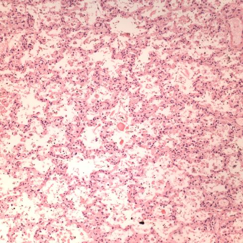
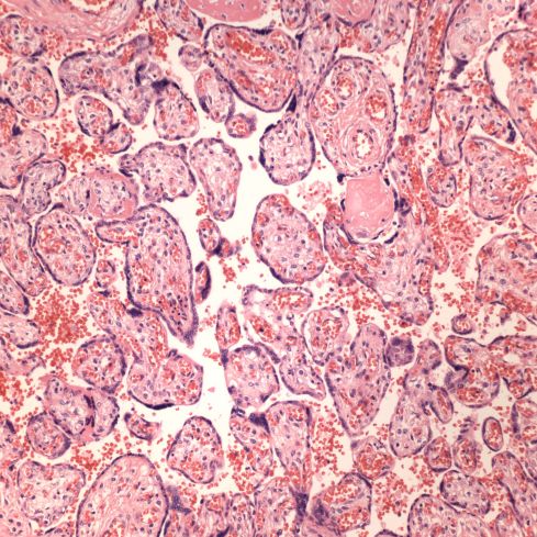
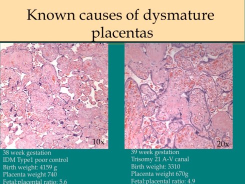
In response to utero-placental ischemia, the placenta will adapt in a way that increases the diffusion of oxygen to the fetus at the expense of nutrient transfer. This change can be looked at as the transition from areas with a more gastrointestinal type function to those with a more lung type function. This process at a less accelerated form underlies the villous changes that we as pathologists see with maturation. The immature placenta has a thick syncytial layer that is metabolically very active with transport. The mature placenta shows many capillary syncytial membranes that minimize the distance between maternal and fetal blood, much like alveolar capillaries in the lung minimize the distance between air and blood. These membranes are associated with trophoblast “knotting”, that is the clumping of apoptotic syncytiotrophoblastic nuclei that are shed into the maternal circulation. This thin barrier between the oxygen source and the capillary bed is needed for efficient oxygen transport. The actual transfer of oxygen is complicated by considerations of the fetal hemoglobin, the overall surface area, the rate and direction of blood flows etc. However, Mayhew’s group have provided a model that shows that decreasing the barrier thickness is the most important adaptation to provide the fetus with sufficient oxygen to survive in a state of decreased utero-placental blood flow6. It comes at the cost of less nutrition and therefore a proportionately smaller placenta and infant, the growth restricted infant.

This very simplified schema of placental insufficiency explains why the premature increase of capillary syncytial membranes and secondarily syncytial knots, the key to identifying utero-placental ischemia under the microscope7. This observation was perhaps first reported by Tenny and Parker, and is sometimes referred to at Tenny Parker change8. The trade off for the increased transfer of oxygen is decreased transfer of nutrients. The villous adaptation has limits, and some infants may develop hypoxia and may even die.

As a practical matter for diagnosis, the lack of villous adaptation to utero-placental ischemia in a growth-restricted infant without other compromising placental lesions is evidence for intrinsic causes of small fetal size.


The maternal systemic response to utero-placental ischemia in preeclampsia


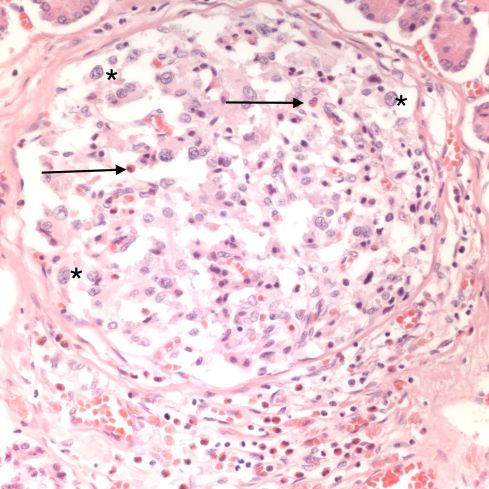
The most common association with utero-placental ischemia based intrauterine growth restriction is severe or early preeclampsia. This is the second response to utero-placental ischemia. Medical students learn that preeclampsia/eclampsia is characterized by edema, gestational hypertension, hyperreflexia (or seizures) and proteinuria with more precise definitions available. Ideally they also learn that this syndrome can be unified as being the consequence of a diffuse endothelial injury. For example, the edema is due to capillary leakage and allows vaso-constrictive blood products to initiate arteriolar hypertension, and damage to glomerular capillaries results in proteinuria. They may also have learned that the cure of preeclampsia is delivery, although some residual effects may persist for weeks. This was the basis for old concept that the placenta was causing a toxemia. That old concept has been justified by molecular techniques that have identified substances from the placenta such as sFLT-1, which compete with endothelial growth factor and interfere with endothelial integrity9,10.

But why would the placenta secrete substances toxic to the mother in the presence of utero-placental ischemia? There is a teleological explanation. First, endothelial leakage allows vasoactive substance to reach the arteriolar smooth muscle causing constriction that increases systemic blood pressure that directly increases perfusion to the placenta. Second, leaking capillaries decrease blood volume increasing the hematocrit and therefore increasing oxygen carrying capacity of the blood. These adaptations may be sufficient to improve oxygen transfer to the infant through a short period of late gestation utero-placental ischemia. They are inadequate to compensate for more severe or earlier onset utero-placental ischemia.

There are still many questions, since other forms of utero-placental ischemia such a maternal thrombophilia with multiple infarctions do not generate preeclampsia. Of course many other forms may not create a large volume of suboptimally perfused villi. The association of a severe early onset preeclampsia with dyandrogenic triploidy suggests a more complex interaction between the placenta and intravillous perfusion than suggested in my admittedly heuristic explanation of improving fetal oxygenation.


Placental infarction


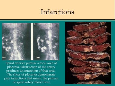
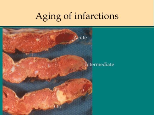
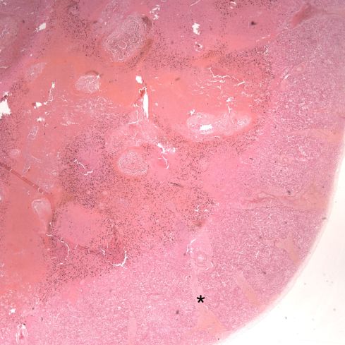
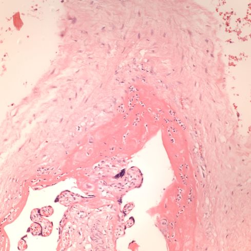
In discussing utero-placental ischemia so far, we have only considered a diffuse loss of placental perfusion. However, a fixed loss of focal perfusion can also have a profound effect on fetal oxygenation. One such lesion is a placental infarction. This is analogous to myocardial infarction in that a permanent occlusion of an end artery (the spiral artery) leads to ischemic death of the supplied tissue. Multiple placental infarctions can significantly reduce the functioning placental volume. Such infarctions occur in some patients with preeclampsia. Typically these infarctions occur at different times as evaluated by the progressive maturation of the infarction11-14. This multi-temporal dimension in preeclampsia may reflect the progressive trophoblast modification of the spiral arteries.

There are two ways that placental infarctions differ from myocardial infarctions because of the circulatory anatomy. First, a placental infarction differs from a myocardial in that the end artery does not occlude a defined capillary bed, but an open intervillous space where flows from adjacent spiral arteries make direct liquid to liquid contact. As we saw on Dr. Ramsey’s injections, the spiral artery flows do not really intermix despite the open space. The concept that each spiral artery has a distinct entrance zone with a central high oxygen flow and a surrounding periphery like the downward cascade of a fountain of lower oxygenated blood has been called a placentone, a persistent unit of spiral artery flow that can be seen looking at the villous morphology of a mature placenta with gradients showing more syncytial knots at the periphery, and larger more intermediate appearing villi in the center15.

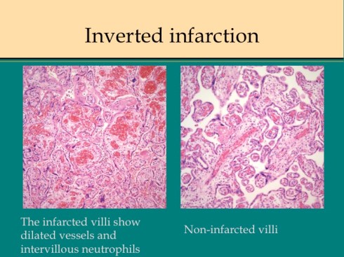
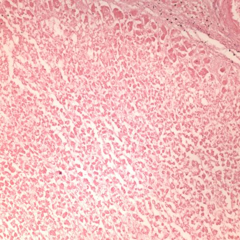
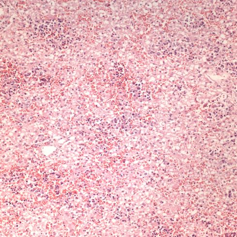
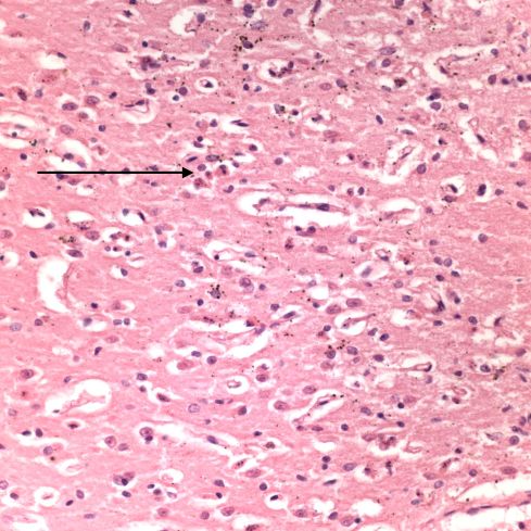
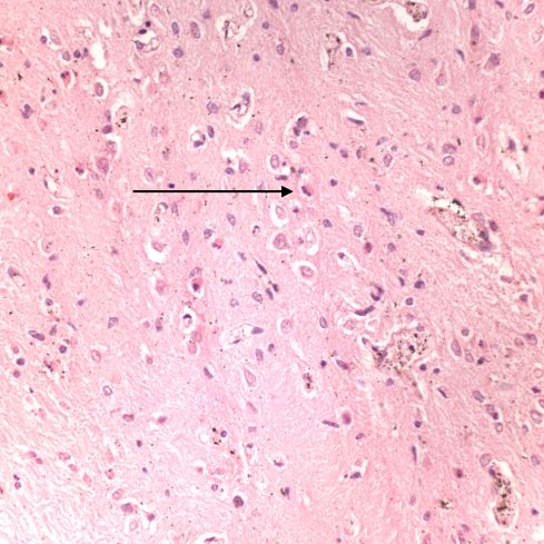
Like a myocardial infarction, an infarction would be expected to allow outside flow into the occluded placentone from collateral circulation, only in the placenta this flow would directly cross the intervillous space. As with myocardial infarctions, placentas with evidence of generalized utero-placental ischemia (analogous to coronary artery disease) tend to have larger areas of infarction than those with normal circulation. The collateral circulation might be expected to create a gradient of hypoxia around the infarction. The histologic observations seem to confirm a gradient. Typically the center shows complete coagulation necrosis of the villi. Initially there is a border of some fibrin and neutrophils like that around a myocardial infarction, but unlike the progress to scar in the myocardium, a shell of perivillous fibrinoid forms around the infarction. This can be understood if 1) the key steps of organization, with ingrowth of capillaries and fibroblasts, can not occur in the intervillous space and villi, and 2) if we accept that lower levels of hypoxia kill syncytiotrophoblast than kill the underlying cytotrophoblastic stem cells. Looking at the histology it would appear that the villous cytotrophoblast not only replenish syncytiotrophoblast but also secrete the components of fibrinoid, such as fetal fibronectin and annexins that make up the extracellular matrix of trophoblast. The hypoxic loss of the syncytium appears to result in fibrin deposition on the bare surface of the villous with stimulation of cytotrophoblast production of matrix. The next layer outward shows marked Tenny Parker changes indicating villous adaptation to hypoxia. Once again I cannot prove this gradient hypothesis, but I can offer it as a way to make sense of what you are seeing under the microscope. Even if truer explanation of placental infarction become available, they will still need to be able to explain what we see under the microscope.

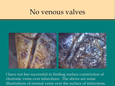
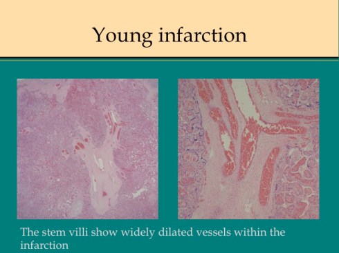
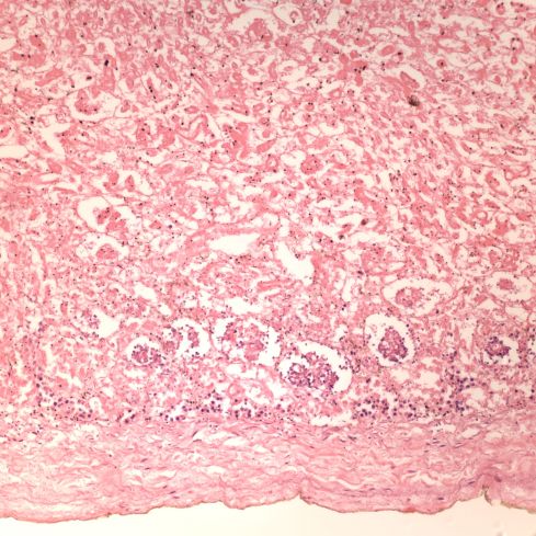
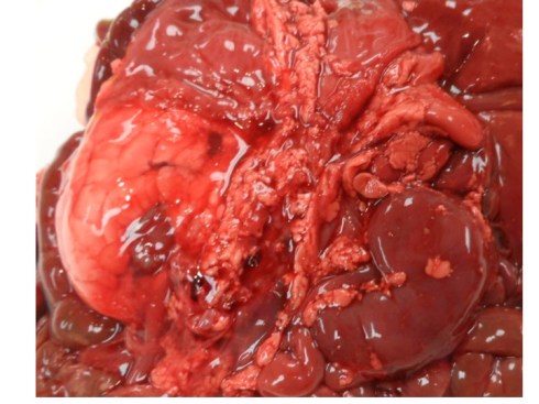
A second difference from myocardial infarction is the dual circulation of the villi. The infarction removes the oxygen supplied by the intervillous circulation, but the fetal circulation of deoxygenated fetal blood is initially still intact. As pathologists we know from observation that early placental infarctions are grossly deep red, and this is due microscopically to dilatation of the fetal vasculature. If we assume that the smooth muscle of the involved fetal vessels becomes hypoxic they might dilate under the fetal blood pressure, in which case the infarcted area would become a low pressure circulatory sink that would not oxygenate the circulation, but add potential harm from thrombogenic material potassium, etc. released from the dying villi. However, the pathologic observations do not support this scenario. There is no accumulation of neutrophils in the fetal vessels that would be expected to be chemotactic to the dying tissue, and thrombi are very rare in the involved fetal vessels. An early obstetrical theory was that placental infarctions were caused by valves closing in the chorionic veins. This theory seems to confuse cause and effect, but something like this may be happening to protect the fetal circulation16. I know of no proof of the following hypothesis, but it seems to fit the pathologic observations.

Like the lung, the efficiency of the placenta would be markedly improved if it could match the fetal circulation to the intervillous circulation, in effect optimizing ventilation to perfusion. Certainly, if the radiologic observations of Elizabeth Ramsey in monkeys are true for the human, spiral artery flow is intermittent, and the placenta would need to efficiently adapt to this variable flow. Constriction of stem vessels would be one way to do this, and indeed these are very muscular vessels, but interestingly unlike vessels in the body there is no clear histologic difference between veins and arteries. If the intervillous circulation became hypoxic, more oxygen might be extracted by slowing the villous flow and dilating the capillaries. This would occur if the villous veins constricted before the arteries. Arterial constriction would raise resistance, and fetal flow would be directed away from the poorly perfused villi. If intervillous flow were to stop as with an infarction or encasement in perivillous fibrinoid, the artery could remain constricted to the point of permanent occlusion. While this is speculation based on the observation of stopped flow and necrosis of the villous vessels in “old” infarctions, the ability of villous arteries to remain constricted has been directly observed in the lamb undergoing placental by pass for experimental cardiac surgery17. When the researchers tried to reattach the placenta to the fetal lamb, the vascular resistance was too high for the lamb to circulate. This appeared to be from arterial constriction due to vasopressin. Likewise, the umbilical arteries constrict and remain so after the cord is cut until the cord becomes necrotic and falls of the umbilicus.

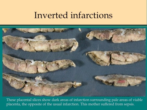
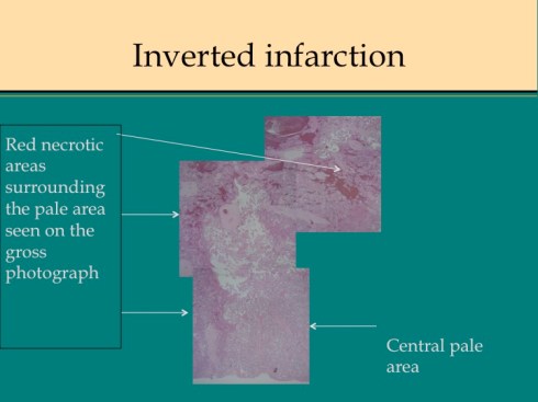
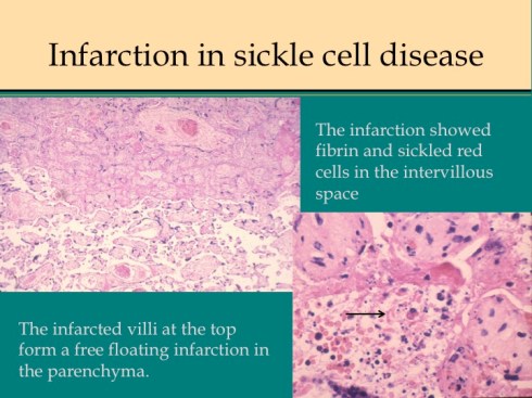
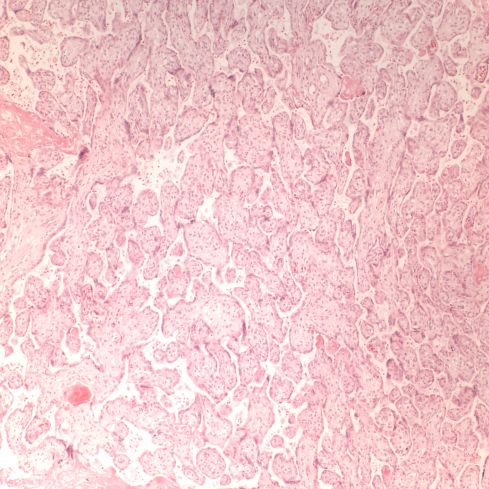
There are some other uncommon causes of infarction. If mother suffers systemic cardiogenic or septic shock, the placenta can become ischemic in such a way that the infarcted villi are at the periphery of flow rather than at the center of the placentone.18 This makes sense since the surround usually shows evidence of greater hypoxia than the center. The placentone centers are typically very pale from not only decreased intervillous flow, but also decreased fetal flow. The peripheral infarction is vasodilated like a typical early infarction. Another infrequent form of infarction is from sickle cell crisis causing thrombosis within the intervillous circulation.


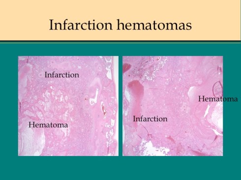
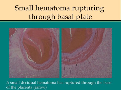
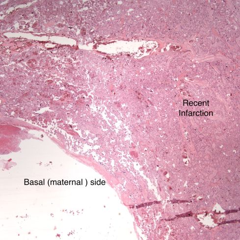
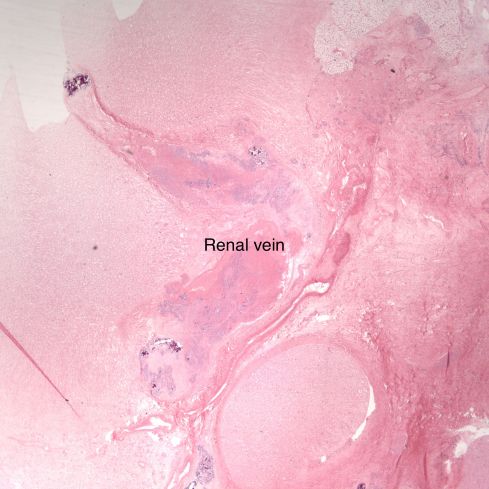
Before we leave the histology of placental infarctions, I want to point out another observation. At the base of even an old “infarction” the basal chorion and attached decidua are viable. This could only happen if the occlusion occurred distal to the separation of the basal endometrial arteries. Direct histological observation of the spiral artery underlying an infarction usually shows just stasis/clotted blood and less frequently any evidence of thrombus. The normal placental histology usually does not allow identification of the cause of the infarction. Perhaps in some cases the infarction is directly due to cellular invasion of the spiral artery or in some cases it may be from conventional fibrin thrombi. Placental bed biopsies have not solved the problem. In a few cases, maternal thrombophilia has been associated with multiple placental infarctions. The spiral artery bed may be more prone to initiating thrombi because of trophoblast remodeling than conventional arterioles adding stasis and endothelial injury to maternal thrombophilia. Some placental infarctions show a central hematoma, which appears to be hemorrhage into an infarction that could be due to either reopening of an occluded spiral artery, or perhaps anastomoses with basal endometrial arteries19.
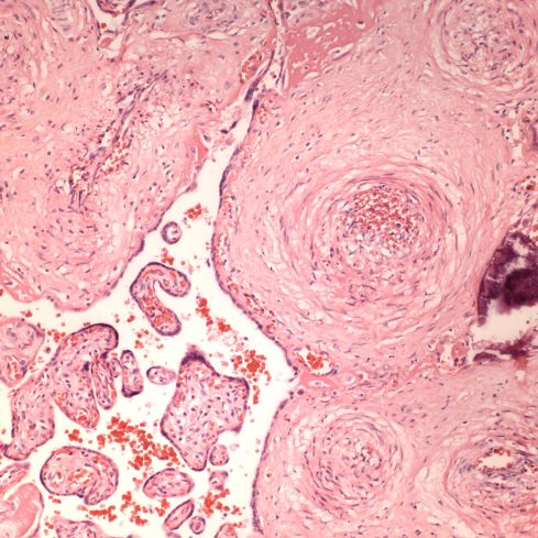
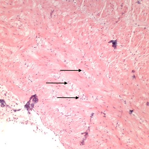
 

Acute atherosis


Uteroplacental ischemia, placental infarctions, and maternal capillary injury are not the only anatomic manifestations of preeclampsia. The spiral arteries not being remodeled by trophoblast, may undergo necrosis with an infiltration of plasma proteins in the wall that cause a fibrinoid appearance and insudation of lipid that results in foam cells in the vessel wall, a lesion termed “acute atherosis”20. Despite widespread endothelial injury in the body with preeclampsia, other arteries do not show this change. Examination of the fetal membranes in woman with preeclampsia show that it is a focal lesion, but appears to be absent in many woman with preeclampsia. Conversely, occasionally the lesion is present in women without any of the clinical criteria of preeclampsia. I don’t know if having the lesion has any long-term significance. Pathologists sometimes confuse the lesion with the changes of trophoblast remodeling. Acute atherosis occurs in non-remodeled arteries in the basal decidua, but the diagnosis is certainly easier in the decidua beneath the reflected fetal membranes.


Placental abruption/ premature placental separation


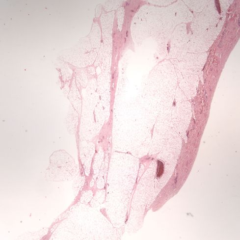
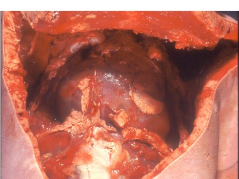
Preeclampsia is the most commonly associated risk factor placental abruption, a term that refers to the clinical presentation of separation of the placenta from the uterus prior to delivery. To understand this concept, consider that the normal mechanism of placental separation is from the shear forces of the uterine contraction after delivery of the infant and amniotic fluid cleave the decidua leaving a basal layer of endometrium in the uterus21,22. The very term decidua reflects this function as in the term deciduous leaves, and if the placenta implants on the myometrium it cannot separate with delivery, hence the term placenta accreta. A premature separation could occur if some force cleaves the decidua. Rupture of membranes in the second trimester results in proportionately more loss of intrauterine volume, and hence more contraction of myometrial area than occurs later in gestation, and may produce sufficient shear to initiate placental separation23,24. Another example of shear stress causing placental separation is the rapid deceleration of an auto accident that results in motion of the placenta in relation to the uterine wall shearing the decidua25,26. Any shearing the decidua results in tearing of the spiral arteries. If the torn area forms a closed space, blood will flow into that space from the spiral arteries until stopped by the intrauterine pressure, forming a retroplacental hematoma. If the decidua can communicate with the cervical os the blood may instead hemorrhage outward. In both cases, the intervillous space is no longer being perfused by the torn spiral arteries and the villi that had been supplied by those arteries will undergo infarction. Usually, more than one spiral artery is involved, and the contiguous infarctions from placental separation are usually larger than an occlusive placental infarction. Smaller separations may not be clinically evident, however, at some point clinical symptoms of placental abruption become evident with uterine pain and tenderness, abnormalities of fetal heart rate tracing, and maternal coagulopathy. From autopsies of stillborn infants with premature separation, a loss of approximately one half of the placenta in a single event is likely to be lethal. Smaller lesions may not have a clinically evident consequence.

Other mechanisms of premature separation likely produce shear by an expanding hematoma in the decidua. In preeclampsia damage to a spiral could produce a hemorrhage that would have the same effect as a spiral artery torn by a shear stress with expansion of the hemorrhage in the decidua potentially shearing adjacent spiral arteries increasing the size of the mass. There is some correlation ironically of abruption with thrombophilia and it is a reasonable pathological concept that in spiral artery thrombi could leads to hemorrhage from basal artery bleeding into necrotic decidua. A similar mechanism may occur with cocaine-induced vasospasm27. Compression of the vena cava elevates uterine venous pressure and hence spiral artery pressures leading to a decidual hematoma28.

In these latter mechanisms, the hematoma appears primary, but with direct shear forces, the hematoma is secondary. The pathology does not reveal the mechanism of a placental separation, but associated infarctions may suggest preeclampsia or thrombophilia as the mechanism.

The pathologist needs to evaluate the extent of a premature placental separation at the gross examination. The placenta may still have an adherent hematoma on the maternal surface. Sometimes only the indentation of the hematoma with a craterous appearance will be found. As the hematoma ages, the color will go from red to brown to tan as hemoglobin becomes deoxidized and leaches out. At the same time the overlying infarction will similarly age. The full extent of the infarcted placenta may not become evident until cut slices of the placenta are looked at. By combining the extent of the compressed intervillous space/visible infarction, the full extent of compromised placenta can be estimated.                        Determining the approximate or relative age of the separation and overlying infarction may also help explain the clinical events. In a very acute clinical abruption, there may be no evidence of an abnormal separation in pathology, even though the obstetrician observed the placenta floating on a hematoma at the time of emergency Cesarean section. By a few hours of age, neutrophils can usually be found within the basal plate of the placenta. The overlying intervillous space is likely to show collapse. The infarction will progress through the same changes as with spiral artery occlusion. The blood clot will gradually become brown and then begin to lose color.

Smaller placental separations can be distinguished from infarctions by the necrosis of the basal plate of the placenta and attached decidua. In general there will also be some hemorrhage within the attached decidua. Intravillous hemorrhage may be increased above an placental separation.

Effect on the fetus


Uteroplacental ischemia leads to adaptation of the placenta that architecturally favors oxygenation over active nutrient absorption. This can eventually result in intrauterine growth retardation. If the utero-placental ischemia becomes more extreme, in theory the adaptive ability of the placenta can be exceeded and the infant becomes hypoxic. This can result in feed forward loop with hypoxia decreasing cardiac efficiency in term increasing fetal tissue hypoxia until fetal acidosis and death occurs. In reviewing stillborn autopsies, I found that stillbirths in growth-restricted infants usually had not just villous adaptation to ischemia but also placental destructive lesions such as infarctions, smaller placental separations or some other lesion such as massive pervillous fibrinoid or massive chronic intervillositis. (see abstract below) A placental separation alone could cause a subacute fetal death if it involved at least 50% of the placenta, assuming no other causes of placental compromise29. Near complete separations not surprisingly showed fetal changes of acute asphyxia. Isolated placental separations of less than 20% demonstrated no long term ill effects30.


  1. Vogel S. Life in Moving Fluids. Princeton, NJ: Princeton University Press; 1994.
  2. Ramsey EM, Donner MW. Placental vasculature and circulation. Philadelphia: W. B. Saunders Company Ltd; 1980.
  3. Whitley GS, Cartwright JE. Cellular and molecular regulation of spiral artery remodelling: lessons from the cardiovascular field. Placenta 2010;31:465-74.
  4. Harris LK. IFPA Gabor Than Award lecture: Transformation of the spiral arteries in human pregnancy: key events in the remodelling timeline. Placenta 2011;32 Suppl 2:S154-8.
  5. Burton GJ, Woods AW, Jauniaux E, Kingdom JC. Rheological and physiological consequences of conversion of the maternal spiral arteries for uteroplacental blood flow during human pregnancy. Placenta 2009;30:473-82.
  6. Mayhew TM, Jackson MR, Haas JD. Microscopical morphology of the human placenta and its effect on oxygen diffusion: a morphometric model. Placenta 1986;7:121-31.
  7. Redline RW, Boyd T, Campbell V, et al. Maternal vascular underperfusion: nosology and reproducibility of placental reaction patterns. Pediatr Dev Pathol 2004;7:237-49.
  8. Tenney Jr. B, Frederic PJ. The placenta in toxemia of pregnancy. Am J Obstet Gynecol 1940;39:1000-5.
  9. Maynard SE, Min JY, Merchan J, et al. Excess placental soluble fms-like tyrosine kinase 1 (sFlt1) may contribute to endothelial dysfunction, hypertension, and proteinuria in preeclampsia. J Clin Invest 2003;111:649-58.
  10. Levine RJ, Lam C, Qian C, et al. Soluble endoglin and other circulating antiangiogenic factors in preeclampsia. N Engl J Med 2006;355:992-1005.
  11. Wallenburg HC, Hutchinson DL, Schuler HM, Stolte LA, Janssens J. The pathogenesis of placental infarction. II. An experimental study in the rhesus monkey placenta. Am J Obstet Gynecol 1973;116:841-6.
  12. Wigglesworth J. Morphological variations in the insufficient placenta. J Obstet Gynecol Br Comwlth 1964;71:87184.
  13. Fox H. The significance of placental infarction in perinatal morbidity and mortality. Biol Neonat 1967;11:87-105.
  14. Wentworth P. Placental infarction and toxemia of pregnancy. Am J Obstet Gynecol 1967;99:318-26.
  15. Schuhmann RA. Placentone structure of the human placenta. Biblthca anat 1982;22:26-57.
  16. Bartholomew RA, Colvin ED, Jr WHG, Fish JS, Lester WM, Galloway WH. Criteria by which toxemia of pregnancy may be diagnosed from unlabeled formalin-fixed placentas. Am J Obstet Gynecol 1961;82:277-90.
  17. Lam CT, Sharma S, Baker RS, et al. Fetal stress response to fetal cardiac surgery. Ann Thorac Surg 2008;85:1719-27.
  18. Bendon RW, Cantor DB. Stillbirth due to placental hypoperfusion after salpingo-oophorectomy for an ovarian cyst. Obstet Gynecol 2007;110:482-4.
  19. Bendon RW. Nosology: infarction hematoma, a placental infarction encasing a hematoma. Hum Pathol 2011.
  20. Khong TY. Acute atherosis in pregnancies complicated by hypertension, small-for- gestational-age infants, and diabetes mellitus. Arch Pathol Lab Med 1991;115:722-5.
  21. Deyer TW, Ashton-Miller JA, Van Baren PM, Pearlman MD. Myometrial contractile strain at uteroplacental separation during parturition. Am J Obstet Gynecol 2000;183:156-9.
  22. Krapp M, Katalinic A, Smrcek J, et al. Study of the third stage of labor by color Doppler sonography. Arch Gynecol Obstet 2003;267:202-4.
  23. Gonen R, Hannah M, Milligan J. Does prolonged preterm premature rupture of the membranes predispose to abruptio placentae. Obstet Gynecol 1989;74:347-50.
  24. Vintzileos A, Campbell W, Nochimson D, Weinbaum P. Preterm premature rupture of the membraes: a risk factor for the development of abruptio placentae. Am J Obstet Gynecol 1987;156:1235-8.
  25. Aitokallio-Tallberg A, Halmesmaki E. Motor vehicle accident during the second or third trimester of pregnancy. Acta Obstet Gynecol Scand 1997;76:313-7.
  26. Rogers FB, Rozycki GS, Osler TM, et al. A multi-institutional study of factors associated with fetal death in injured pregnant patients. Arch Surg 1999;134:1274-7.
  27. Acker D, Sachs BP, Tracey KJ, Wise WE. Abruptio placentae associated with cocaine use. Am J Obstet Gynecol 1983;146:220-1.
  28. Mengert WF, Goodson JH, Campbell RG, Haynes DM. Observations on the pathogenesis of premature separation of the normally implanted placenta. Am J Obstet Gynecol 1953;66:1104-12.
  29. Bendon RW. Review of autopsies of stillborn infants with retroplacental hematoma or hemorrhage. Pediatr Dev Pathol 2011;14:10-5.
  30. Bendon RW, Coventry S, Bendon J, Nordmann A, Schikler K. A follow-up study of lympho-histiocytic villitis and of incidental retroplacental hematoma. Pediatr Dev Pathol 2014;17:94-101.


911Slide1104514151920212930

Subutex modification of placental maturation?

23 Feb

In the course of performing pathologic examination of placentas, our group noted an increase in the diagnosis of villous dysmaturity in placentas from patients being treated with Subutex for heroin addiction. We internally tested our ability to recognize the villous dysmaturity in these patients by comparing two sets of 4 placentas respectively from mothers treated with Subutex or Methadone therapy. Blinded to the clinical history, our group of pathologists consistently identified the slides from the mothers with Subutex as showing villous dysmaturity and no abnormality in the methadone group. This observation was presented at the Society for Pediatric Pathology perinatal slide review in Toronto. (see attached Powerpoint slides). At the presentation, Dr. Fusun Gundogan from the Women and Infant Hospital of Rhode Island confirmed making a similar observation. We sent this pathologist our 2 sets of test slides without any clinical history, and she also recognized villous dysmaturity on 3 of the 4 placentas from the Subutex treatment placentas and none from the methadone treated placentas.

Subutex has recently replaced methadone as the treatment for heroin addiction in pregnancy because of studies showing less neonatal morbidity1-3. The active drug is buprenorphine and there are variable dosing schedules. It crosses the placenta4.

Delayed villous maturation is a somewhat subjective placental diagnosis that in our practice refers to an increased number of immature villi (large, irregular shape, with thick even syncytiotrophblast and few syncytial knots) compared to normal maturation5. The diagnosis is most often evident with aneuploidy particularly trisomy 21, and rarely with diabetes unless there is very poor glucose control. In many placentas with the diagnosis, there is no immediate clinical correlation.

The villi in the placenta mature to increase the surface area for oxygen diffusion. The rate of oxygen diffusion from maternal to fetal blood is inversely related to the third power of the barrier thickness6. Therefore maximum oxygen exchange occurs in villous areas of capillary syncytial membranes in which the fetal capillary is at the surface, and the syncytiotrophoblast is markedly attenuated. The development of such membranes is associated with the clumping of apoptotic syncytial nuclei in adjacent areas. As demonstrated by radiographic cinematography in the primate placenta, the intervillous maternal blood enters from the spiral artery in a fountain effect7. This area of perfusion from a single spiral artery has been termed the placentone8. Typically, the periphery of the flow is the first area to show placental maturation, most easily seen as an increase in syncytial knots.

Larger, immature villi have more blood vessels in the center of the villi. A placenta with fewer mature villi would be expected to be less efficient at oxygen exchange. One hypothesis for the persistent immature villi is that they are an adaptation to better intervillous blood flow such as having slower, high volume flow with better oxygen transfer. The other explanation is that there is a pathologic delay in maturation or a misreading of the normal signals that induce maturation. If the first explanation is true the infants should have normal labors because the fetus can recover oxygenation between contractions. If the second explanation is true, then some abnormalities of labor would be expected.

Women with a history of heroin addiction may be more likely than the average population to have used other drugs including alcohol and cigarettes. To test any hypothesis in this group of patients, the ideal controls would be matched for this difference from the general population. A proxy for such matching is to compare historical controls having methadone treatment. There is also a need to recognize that patient reported use of drugs and medications including Subutex may not reflect actual use.

I had considered trying to coordinate a larger study to confirm this possible association of villous dysmaturity and Subutex, but it proved more complex than I could undertake. I would be interested if any one else has made a similar observation.


References:


  1. Kakko J, Heilig M, Sarman I. Buprenorphine and methadone treatment of opiate dependence during pregnancy: comparison of fetal growth and neonatal outcomes in two consecutive case series. Drug Alcohol Depend 2008;96:69-78.
  2. Jones HE, Kaltenbach K, Heil SH, et al. Neonatal abstinence syndrome after methadone or buprenorphine exposure. N Engl J Med 2010;363:2320-31.
  3. Brogly SB, Saia KA, Walley AY, Du HM, Sebastiani P. Prenatal buprenorphine versus methadone exposure and neonatal outcomes: systematic review and meta-analysis. Am J Epidemiol 2014;180:673-86.
  4. Concheiro M, Jones HE, Johnson RE, Choo R, Shakleya DM, Huestis MA. Maternal buprenorphine dose, placenta buprenorphine, and metabolite concentrations and neonatal outcomes. Ther Drug Monit 2010;32:206-15.
  5. Redline RW. Classification of placental lesions. Am J Obstet Gynecol 2015;213:S21-8.
  6. Mayhew TM, Jackson MR, Haas JD. Microscopical morphology of the human placenta and its effect on oxygen diffusion: a morphometric model. Placenta 1986;7:121-31.
  7. Ramsey EM, Donner MW. Placental vasculature and circulation. Philadelphia: W. B. Saunders Company Ltd; 1980;22:26-57.
  8. Schuhmann RA. Placentone structure of the human placenta. Biblthca anat 1982;Slide5.jpgSlide1
  9. Slide2Slide3Slide4.jpg


A study of stillbirth

26 Jan

Below is a rough draft of a study idea that I had hoped would make real progress on understanding the causes of stillbirth. It addresses only those deaths due to asphyxia, but from my autopsy experience this is likely to be a large number of deaths. The proposal is somewhat under-referenced, but the pages I have been adding to this site will address the appropriate literature review. I can not realistically undertake this project, but I hope the ideas might at least be useful to others interested in the topic.

Aims:

  1. To demonstrate the mechanism of asphyxia in stillbirth by investigating the intrauterine ultrasound and the maternal history in detail at the time of discovery of the intrauterine death.
  2. To correlate the above findings with the autopsy and placental examination in light of the above clinical investigation
  3. To use the information from aims 1 and 2 to develop hypotheses for potential prevention of stillbirth


Background:


There remains a group of stillborn infants that even a careful clinical, autopsy, and placental investigation fails to find the cause. This is particularly frustrating in appropriate for gestation infants without malformations, genetic disease, fetal hydrops, or infection. For analysis, this group of cases can be separated from infants born prior to extra-uterine viability in which prevention of stillbirth would be achieved by preventing very early preterm labor. The autopsy of these unexplained stillborn infants often shows evidence of the mechanism of death that is consistent with either very acute asphyxia or rapid onset of heart failure presumably from hypoxia/acidosis (see below).

The evidence of acute asphyxia is the finding of intrathoracic petechiae, much like those found in SIDS. These petechiae can be formed experimentally by tracheal occlusion at end expiration, and this is likely the mechanism in SIDS in that the infant exhales and then initiation of inhalation is blocked by a physical object. In utero, asphyxia is the stimulus to start respiration and occlusion may be a glottic stop mechanism such as that proposed to assist the first attempt of ventilation after birth. Intrathoracic petechiae are found with large placental abruption, an anatomically observable mechanism of total or near total acute asphyxia documented by placental examination(1). From experience they are present in other cases with a clear mechanism of relatively rapid asphyxia such as umbilical cord hematoma. Acute birth asphyxia as demonstrated by Dawes(2) and others results in fetal gasping that if the airway were occluded would produce negative intrathoracic pressure that could expand surface capillaries and result in their rupture. In our preliminary data such petechiae are present in 15% of all stillbirths greater than 23 weeks of gestation.

In the same study of abruption, placental separations that were small but still lethal demonstrated modest pleural and pericardial effusions, and dilated cardiac chambers in the infant. I hypothesized that this was due to acute heart failure from hypoxia leading to a positive feedback loop of hypoxia decreasing cardiac output, which decreases oxygenation, which decreases cardiac output further. In general more chronic causes of fetal hydrops produce more prominent edema, and lung hypoplasia. Like intrathoracic petechiae this pattern is also present in stillbirths without abruption, some with explained asphyxia and some with no anatomic cause. This pattern appears to be more common in small for gestation infants especially those with fixed anatomic loss of placental functional volume.

The unexplained stillborn autopsies with evidence of asphyxia producing either gasping or heart failure likely have an anatomic cause. Many potential mechanisms would be very difficult to detect by examination of the placenta or infant after delivery. For example we demonstrated in vitro that if wrapping of the cord leaves only a short free segment, flow in the umbilical vein can be stopped by modest torsion of the cord(3). In vivo this would correspond to a cord wrapping that resulted in a short segment between the umbilical insertion and the beginning of the wrapping. Aside from twisting, direct stretching of the helical structure of the cord might also interfere with blood flow in the short segment. In other cases, the position of the cord in relation to the presenting part and pelvis might lead to cord compression from occult prolapse or undetected funis previa. Oligohydramnios may favor the cord being trapped, and this would not be detectable after delivery. Other anatomically “invisible” factors such as maternal ketosis, hypoxia, sleep apnea, decreased uterine flow for example from venal caval compression with supine position could be contributing factors to fetal hypoxia. Some of the factors may be detectable by directed ultrasound at the time of fetal death, while others may be detectable by a detailed and open-ended maternal history and examination.

Our goal is to not use a risk factor approach, but rather a deductive approach to solve the cause of death in the individual infant. Obesity is a risk factor for stillbirth(4), but we want to discover the mechanism of death of a particular infant in that obese mother. The hope is that some of those mechanisms may be detectable and preventable without the difficulty of reducing the overall risk factor, for example obesity. Our approach will be sensitive and reassuring to the parents’ anxiety and guilt, and will attempt to secure their involvement in the process of discovering the unknown mechanisms of stillbirth. Our approach also involves a detailed cooperation of clinician and pathologist in a mutually informing interaction in interpreting all of the findings in individual cases, and then pooling the information. This study would obtain detailed information on traditionally known and unknown causes of stillbirth.


Methods:


Any pregnancy with intrauterine fetal death is eligible to be enrolled in the study. The triggering event is the detection of the death, and immediate notification of a study member on call to request consent and begin the process. The first step after consent is a detailed ultrasound noting fetal position, amount of amniotic fluid, any structural details of the placenta, the distance of free umbilical cord from the placental insertion if that can be measured, and inspection of areas around the fetal presenting part in the pelvis for evidence of umbilical cord compression. A discussion with the parents is initially open ended as to events in the previous week, day, hours with directed questions about fetal movement, use of medications, trauma, with the approach of encouraging the parents to talk about their own concerns, and to record this information. We will speak with parents separately and together. A normal routine review of systems by history and exam, and review of the prenatal record and prenatal tests and ultrasound would be included. The physical exam would include cervical dilatation, maternal weight and height compared to pre-pregnancy, and vital signs. Maternal blood would be drawn for possible infection titers, K-B, and saved serum for potential markers and DNA. A EDTA tube would be drawn for possible coagulation markers. The goal is to obtain the specimen but the testing would be determined on algorithms based on history, and findings in the placenta and the fetal autopsy. The consent would include permission for placental examination. The autopsy consent would follow the normal procedures including the extent of the autopsy, and the disposition of the infant. To the extent possible, the obstetrician and other clinical caretakers of the infant will be included in all the consent process as additional resources for the patient’s questions. If the membranes have not ruptures amniotic fluid will be obtained.

The examination of the placenta starts with measuring the entire cord length on the infant and on the placenta. The cord and placenta are photographed with fetal and maternal surface, and two longitudinal photographs of the cord. A standard pathology protocol would be followed that records umbilical cord: vessel number, insertions, color, average diameter, and lesions. Membranes: color, rupture, and lesions. Fetal surface: margin, color, vessels, lesions, Maternal surface: completeness, lesions and cut sections at 1 cm intervals but also cuts of all palpable lesions. The cute sections are also photographed. Routine sections are taken (ends and middle of the umbilical cord, rupture edge to margin of the fetal membranes, and at least 3 sections of the placenta including one through the umbilical cord insertion, as well as sections of lesions as needed). A sample of fresh rinsed placental villi and cord will be frozen for storage as well.

The stillborn autopsy will also follow a specific protocol. The external examination will include the usual measures and body weight. The examination will include the physical portion of the Ballard score to estimate gestation with documenting photographs. The photographs will also be taken to document the extent of skin slippage, fetal hemoglobin staining of the cord and sclera, and the fullness of the fetal skull. A radiograph will be taken AP and lateral if possible with a Faxitron or fine resolution to detect opacities in the fetal liver and bowel. The internal examination will include a window in the chest to measure and obtain pleural fluid volume and sample, a lung culture, and a photograph of the anterior thoracic cavity before dissection and again after removal of the entire thymus and pericardium. A separate photograph will be made of the ductus arteriosus in situ after removing the vagal bundle for visibility and retracting the left lung. A photograph of the abdomen in situ will also be made to judge the liver size, color and the color of the colon. A routine autopsy and sampling will be done by protocol adaptable to the findings as well as those of the history and placental findings. A portion of fetal liver and brain will be frozen. The routine heart examination in a fetus usually follows the dissection along the lines of blood flow. If the heart appears externally normal based on a systematic examination e.g. symmetry of the great vessels, symmetry of the ventricle, location of the great vessels and pulmonary veins, etc ) then the heart will be fixed and cut in cross section in the coronal plane of the posterior atrio-ventricular junction at 3 mm intervals and photographed and measured to determine dilatation. Sections will be made to include the tips of the papillary muscles to look for myocardial necrosis.

One aim of the placental and autopsy examination is to develop the most useful protocol for evaluating the mechanism of stillbirth. The baseline protocols recognize the value of current practice, but add some additional information, and are designed to retain photographic and physical materials for reevaluation. The baseline information obtained will be entered into a Filemaker Pro database that allows for easy modification, multiple screens for different types of information.

The process is one of collaboration and critical review, and ongoing modification. Each autopsy is reviewed within in the institution and at least one of the co-PI in both pathology and obstetrics and a critique produced. The findings are presented to the parent(s) in a conference. The parent’s questions and further information are also entered as data. All of the cases are reviewed monthly from the Filemaker database to monitor the success of collection of data, to identify potential improvements. Annually, all the cases are reviewed by all co-PI’s and ideas are presented at an annual meeting.

Hypothetical examples of the conference: If there is evidence that unusual physical exertion or weight loss, or gastrointestinal illness preceded stillbirth in obese patients, does the autopsy confirm fetal heart failure possible related to ketosis. Is there published information on models of this effect? Are there experts in this area that we can consult? Can we quantify myocardial glycogen in the infant, and do we know the effect of intrauterine postmortem retention time on those levels. Do we need to freeze a myocardial sample on cases for either frozen section PAS stain or direct assay of glycogen levels. How many cases that might relate to this cause are likely to occur annually. Are there better ways to evaluate maternal blood or urine? Should we collect a urine sample on obese mothers in the study? How important is underlying maternal glucose intolerance? Should we also test HA1c in these cases? How many more stillbirths would be needed to recruit to test the hypothesis of ketosis as a mechanism of death. The uniqueness of this process is that the direction of the study cannot be anticipated since it develops from the data being acquired. It may be that obsese patients have increased stillbirth because of pelvic narrowing from internal adipose with increased chance of umbilical cord entrapment against the presenting part, an insight that would trigger an entirely different approach to further the research into mechanism, risk and prevention.

The same approach would be applied to known mechanisms of death such as placental abruption. In those cases, even with known risk factors such as preeclampsia, cocaine, or thrombophilia, there may be common historical factors such as prolonged supine position or physical deceleration that might turn a potential into an actual retroplacental hematoma, or there may be better predictors such as other small retroplacental or retromembrane hematomas in context with the other risk factor. Even if there are no indirect clues, more systematic evaluation of the fine structure of the retroplacental hematoma microscopically might pin point the area of initiation and growth of the separation to better understand dynamics and possibly the timing. The question to be answered is why did this particular patient have an abruption and can that provide a more general insight in understanding the mechanism and preventing it

The only inclusion criterion is a prenatally detected stillbirth. Ideally recruiting a core of four or five institutions with more than 10,000 deliveries per year should provide 80 to 100 cases per year for inclusion. Part of the goal of this methodology is to have a core of committed people becoming increasing knowledgeable about stillbirth. Each institution would have two main Co-PIs one in maternal fetal medicine or obstetrics, and one in pathology, and care-giver teams.

There is no statistical analysis for this study. The approach is unconventional, and the outcome is the production of new testable hypotheses and creation of a core of experienced researchers in this area. There is also an expectation that the hypothesis about stillbirth will also have bearing on prepartum and intrapartum asphyxia that can cause neurologic injury to the infant.


References:


  1. Bendon RW. Review of autopsies of stillborn infants with retroplacental hematoma or hemorrhage. Pediatr Dev Pathol. 2011;14(1):10-5.
  2. Dawes G. Foetal and Neonatal Physiology. Chicago, IL: Year Book Medical Publishers, Inc.; 1968. 247 p.
  3. Bendon RW, Brown SP, Ross MG. In vitro umbilical cord wrapping and torsion: possible cause of umbilical blood flow occlusion. J Matern Fetal Neonatal Med. 2014;27(14):1462-4.
  4. Yao R, Ananth CV, Park BY, Pereira L, Plante LA, Perinatal Research C. Obesity and the risk of stillbirth: a population-based cohort study. Am J Obstet Gynecol. 2014;210(5):457 e1-9.